Orthopedics - Oleksa A.P. 2006
Congenital malformations of the upper extremity
Congenital malformations of the forearm
Madelung's disease
Madelung's disease, or rather Madelung's deformity, is named after the author who in 1878 described this characteristic deformity in the wrist joint area. It most commonly occurs in children aged 13–15, but can also be seen in younger children as well as in older individuals aged 37–39 (Lordkipanidze E.F., 1957).
Madelung's deformity is typically bilateral (80%), occurs four times more frequently in girls than in boys, and develops gradually and imperceptibly during Puberty (Ioffe A.A., 1936; Shyriak F.M., 1963).
Class="center">
Fig. 117. Radiograph of Madelung's deformity (wrist joint in two projections).
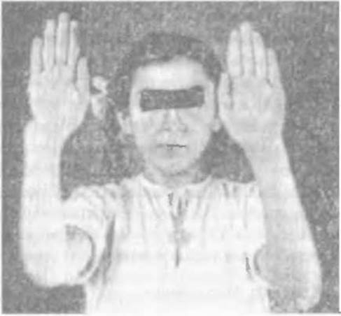
Fig. 118. Clinical presentation of Madelung's deformity of the right hand (Trubnikov V.F.).
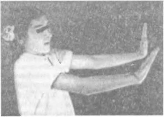
Fig. 119. Limitation of wrist extension.
The classic clinical manifestation of the deformity is a bayonet-like shape of the wrist joint region, with a pronounced protrusion of the ulnar HEAD and volar flexion of the distal end of the radius. Consequently, the hand is inclined toward the ulnar and volar sides, somewhat resembling Clubhand (Fig. 117). This anatomical bone alignment causes significant restriction not only in flexion-extension Movements of the hand, but also in supination-pronation.
Difficult wrist extension is caused by the excessive protrusion of the dorsal edge of the volarly flexed distal end of the radius (Fig. 118), while rotational movements are sharply restricted or even impossible due to the disruption and mismatch of the articular surfaces in the distal radioulnar joint (Fig. 119).
There are several theories regarding the causes of the deformity, including the dominance of flexor Muscles over extensors (Madelung), trauma, Rickets, etc. The pathogenetic essence of the deformity lies in the curvature of the distal end of the radius resulting from damage to its metaepiphyseal region (Petukhova L.I., Vasershtein I.S., 1961), primarily the ulnar region of the radial epiphyseal Cartilage (Notoveltnov S.A., 1926; Scharizer E., 1956).
However, cases have been described (Polyak L.S., 1929) of excessive curvature of the radial diaphysis in the palmar and ulnar directions. Occasionally, albeit very rarely, medial Torsion of the radius may occur, which positions its styloid process in such a way that the radius appears shortened and the ulna longer (Springer C., 1911). During the child's growth, this deformity affects the characteristic arrangement of the proximal carpal row. Instead of an arched formation, the bones form a wedge with the lunate bone at its apex. Furthermore, this cannot be considered a dislocation in the wrist joint, because even in severe cases of Madelung's deformity, the articular surfaces remain closely approximated and maintain near-normal relationships.
Treatment. The practical experience of clinicians indicates that conservative treatment for Madelung's deformity is ineffective. Massages, physical therapy, manual manipulations, and corrective splints do not help patients and are therefore no longer used. Among the proposed surgical Procedures to correct the deformity, corrective osteotomy of the radius combined with ulnar resection remains the most justified, the technique of which was developed at the Sytenko Kharkiv Research Institute of Orthopedics and Traumatology.
Initially, under anesthesia, a wedge osteotomy is performed in the epiphyseal region of the radius, ensuring that the Base of the wedge is positioned on the radial and dorsal sides of the bone, followed by resection of the distal part of the ulna. The width of the wedge base is calculated prior to surgery using radiographs in two projections. To do this, osteotomy lines are drawn on the images in pencil to determine the shape of the wedge, or a paper template of the bone is cut out along these lines, followed by a paper wedge of the required shape.
Precise preoperative planning prevents potential errors, as the wedge osteotomy of the radius and the extent of ulnar resection are closely interrelated; the final anatomical, cosmetic, and functional outcomes of the surgery depend on this (Fig. 120).
The fragments of the osteotomized bones are perfectly repositioned and fixed with metal implants. Following surgery, an immobilizing plaster splint is applied to the arm for three weeks, after which rehabilitation therapy is initiated.
Good results have also been achieved by Polyak L.S. (1929), Kholevich Ya. (1957), and others following wedge osteotomy of the radius and resection of the ulnar head with division of the pronator quadratus Muscle, as well as after hinge osteotomy of the radius and oblique osteotomy of the middle third of the ulna with division of the pronator teres.
As a rule, surgery is performed on adults after growth has ceased, since Madelung's deformity manifests in adolescence and progresses slowly.
Mention should also be made of the "anti-Madelung" deformity, which occurs as a complication (Fig. 121) in chondrodystrophy and multiple cartilaginous exostoses. The Essence of this deformity consists of shortening of the ulna (ulnar clubhand), in contrast to Madelung's deformity, where the radius appears shorter due to its distal bending.

Fig. 120. Diagram of wedge corrective resection of the radius and ulna in Madelung's deformity.

Fig. 121. Anti-Madelung deformity in chondrodystrophy (Reinberg S.A., 1964).
Last update: 10/08/2026
Editorial and Educational Adaptation: This material has been compiled based on the primary/original source text. The project team performed an editorial review, corrected technical inaccuracies, structured sections, and adapted the content for an educational format.
What was processed:
- elimination of formatting defects (OCR errors, structural breaks, corrupted characters);
- editorial organization of content;
- standardization of terminology in accordance with academic sources;
- verification of factual statements against the original source text.
All mentions of the author, publication year, and origin of the primary text have been preserved in accordance with the source.