Orthopedics - Oleksa A.P. 2006
Neurogenic Deformities of the Musculoskeletal System
Paralytic Clubfoot
Paralytic Clubfoot occurs due to paralysis of the m. peroneus longus and brevis, sometimes combined with paralysis of the toe extensors. It is the most common clinical form of paralytic FOOT deformity, accounting for 39.7% (Vreden R.R.) to 56% (Friedland M.) of cases.
Due to the tension of the Muscles that have retained their innervation, pes equinovarus develops. The severity of the deformity varies. In mild cases, the talus retains its anatomical shape and fits properly into the mortise of the ankle joint.
In more severe cases, as the patient grows, the talus changes shape, becoming wedge-shaped and subluxated. If the child walks, pressure is transmitted to the navicular bone, which also becomes wedge-shaped. Foot supination is caused by the tension of the calcaneus from the triceps surae Muscle (m. triceps surae). As growth continues, the cuboid bone enlarges and becomes quadrilateral. Over time, this clubfoot becomes fixed because, alongside the bony changes, soft tissue alterations occur: the Tissues on the medial side of the foot become coarse and shortened, while the joint ligaments and plantar aponeurosis become tight, holding the foot in this position.
When walking, the patient bears weight on the lateral edge of the foot, leading to The formation of a classic corn and Skin callosity.
Treating paralytic clubfoot is quite challenging and depends directly on the severity of the deformity.
If the patient has received regular Treatment involving massage, passive joint mobilization, corrective plaster splints, and orthopedic footwear, the clubfoot is easier to correct. In neglected cases with pronounced paralytic clubfoot, preliminary preoperative conservative management is required. Its goal is to correct the existing foot deformity through progressive stretching of the contracted muscles and joint ligamentous apparatus, reduction of subluxations, etc. Following each stage of manipulation (redression), a short leg cast (boot cast) is applied to maintain the corrected position of the foot. Redresgements with cast changes are performed every 7–10 days.
For mild forms of clubfoot, two to three redressions are usually sufficient to correct the deformity, whereas severe cases require a longer preparation period before surgery. If it is not possible to bring the foot into the desired position, the surgical plan must be modified.
Once the Components of the clubfoot have been corrected conservatively, this foot position must be stabilized by muscle transposition. Five types of surgical interventions have been proposed for this purpose.
1. Most commonly, tendon transposition of the tibialis anterior muscle (m. tibialis ant.) is performed, which ranks second in strength only to the m. gastrocnemius. Converting this muscle from a supinator into a pronator significantly reduces supination and adduction of the distal part of the foot (Fig. 374).
As a rule, supination and adduction are combined with foot flexion (equinus) due to weakness of the extensor digitorum communis muscle.
Nikiforova E.K. (1968) suggests beginning the surgery with a Z-plasty incision and lengthening of the triceps surae muscle (m. triceps surae), followed by transposition of the tibialis anterior tendon according to Vreden V.V.
In cases of pronounced foot excavation, the surgery is additionally supplemented by a transverse incision of the plantar aponeurosis (fascia plantaris).
Surgical technique. Under anesthesia and using a tourniquet, an oblique incision of the skin and superficial fascia is made along the Achilles tendon so that the postoperative scar does not lie over the prominent area of the tendon. After exposing the tendon, a Z-lengthening is performed in the sagittal plane so that the distal segment remains attached to the calcaneal tuberosity. The foot is manipulated and brought into hyperextension, and the tendon is sutured with the foot positioned at a 90° angle relative to the lower leg.
To transpose the m. tibialis ant., three incisions are required. First, the insertion site of this muscle tendon is exposed and detached through an initial incision, and then delivered into the wound via an incision on the anterior surface of the lower leg lateral to the crista tibiae at the musculotendinous junction. Next, a third incision is made over the PROJECTION OF THE fifth metatarsal bone and connected subcutaneously using a hemostat (cornutong) to the incision on the lower leg. The tendon is passed through this tunnel and secured into an osteoperiosteal bridge on the fifth metatarsal bone so that the foot is at a 90° angle to the lower leg and slightly pronated. The tendon must run straight through the tunnel without kinking, underneath the inferior extensor retinaculum of the toes.
The wounds are closed in layers. A short leg cast is applied in the same foot position for 4–5 weeks. Afterward, the patient must wear orthopedic footwear for a year.
Class="center">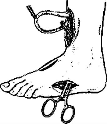
Fig. 374. Schematic drawing of the tibialis anterior tendon transposition to the lateral border of the foot.
2. If the deformity in paralytic clubfoot is not very pronounced and the function of the toe extensors, the extensor hallucis longus, and the triceps surae is preserved, Sachenko A.G., Frumina A.Ye., and others recommend transposing the tendon of the tibialis posterior muscle (m. tibialis post.). This muscle is strong enough to reduce supination and equinus.
The mobilized tibialis posterior tendon is transposed by passing it through a tunnel created with a hemostat through the interosseous space and attaching it subperiosteally to the cuboid bone.
Nikiforova E.K. routes this tendon anterior to the Achilles tendon into a wound on the lateral aspect of the lower leg and fixes it to the cuboid bone beneath an osteoperiosteal bridge. In both variants, the transposed muscle fairly quickly restores its function.
If a mild form of clubfoot is combined with an equinus deformity, the tendon must be lengthened following a Z-plasty incision, and the foot is set in slight overcorrection.
Following surgery, a standard cast is applied from the tips of the toes to the middle third of the thigh. After 10 days, the knee is freed to restore joint mobility. At four weeks, the cast is removed, and rehabilitation therapy is initiated. The patient must use orthopedic footwear for at least a year.
3. Regarding the transposition of the extensor hallucis longus tendon (m. extensor hallucis longus), Frumina, Nikiforova, Carpenter, and others believe that transposing it alone (as an isolated Procedure) is ineffective, except when combined with other stabilizing Procedures or the transposition of other muscles in paralytic clubfoot.
4. There are also opponents and proponents of transposing a portion of the triceps surae tendon. Nikiforova argues that this surgery does not restore foot function because a single muscle cannot simultaneously act as a flexor, pronator, and extensor of the foot.
However, Akatov M.V. and Itskova M.A. suggest performing a split triceps transfer, arguing that the gastrocnemius and soleus muscles are connected only by their tendons, while their muscle bellies have separate Blood supplies, innervation, and attachment points.
Surgical technique according to Itskova M.L. (1950). Under anesthesia and using a tourniquet, an incision is made through the skin, subcutaneous tissue, and fascia along the posterior aspect of the leg, extending from the border of the middle and upper thirds down to the heel. The exposed m. gastrocnemius is bluntly separated from the m. soleus in a downward direction, and their interconnected tendons are split frontally with a scalpel all the way down to the calcaneal tuberosity. Next, the tendon of the m. gastrocnemius is detached from the calcaneal tuberosity along with a small bone fragment for elongation, and passed through a subcutaneous tunnel into the area of a second incision made on the dorsal aspect of the foot. This tendon is fixed periosteally to the Base of the Metatarsal Bones. If pes aequinus is present, the Achilles tendon is lengthened via a Z-plasty incision.
Following surgery, a plaster cast is applied and removed after three weeks. Patients are required to wear orthopedic footwear for one year.
This procedure is technically demanding and is therefore performed by a limited number of specialists.
Itskova M.A. pointed out that achieving independent function of the separated muscles is difficult, and their functional recovery is slow and prolonged.
Movshovich I.A. proposed the separate transposition of the heads of the triceps surae muscle (surgical technique according to Movshovich I.A. (1956), Fig. 375). Under anesthesia, a longitudinal incision is made along the posterior aspect of the leg, starting from the level of the fibular HEAD downwards and lateral to the Achilles tendon, extending onto the dorsolateral surface of the foot to the insertion site of the peroneus brevis tendon. After exposing the muscles, the m. gastrocnemius and m. soleus are bluntly separated using an elevator, and the Achilles tendon is split frontally down to the calcaneal tuberosity. Subsequently, the m. gastrocnemius and its tendon are divided into two halves. The tendon of the fibular muscle is then routed anterior to the lateral malleolus and sutured to the tendon of the lateral head of the m. gastrocnemius.
If the tendon of the lateral head can be pulled down to the lateral border of the foot, the peroneal tendon does not need to be disturbed. If necessary, a Z-lengthening of the m. soleus tendon is additionally performed. Afterwards, an incision on the anterior aspect of the lower third of the leg exposes the tendon of the m. tibialis anterior, to which the tendon of the medial head of the m. gastrocnemius is sutured, having been passed through a subcutaneous channel from the medial side of the leg.
If the tendon of the medial head can be pulled down to the first cuneiform bone, it is fixed directly to it.
After the operation, a plaster cast is applied for five weeks, followed by a comprehensive rehabilitation program.
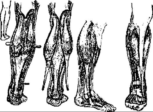
Fig. 375. Schematic diagram of Movshovich's surgical procedure.
Special attention should be paid to therapeutic physical culture (TPC), which contributes most significantly to functional recovery.
5. Combined surgeries involve the transposition of the tendon insertions of muscles that have retained their innervation, combined with stabilizing procedures on joints and bones. If the pronators and extensors of the foot are paralyzed, optimal results can be achieved by combining muscle transposition with a triple arthrodesis (between the talus and calcaneus; calcaneus and cuboid; and talus and navicular bones).
The surgery is performed under general anesthesia. First, the Achilles tendon is lengthened via a Z-plasty incision, followed by the transposition of the tibialis anterior tendon using the classic Vreden technique (three incisions). Utilizing two incisions on the foot, a triple arthrodesis is performed, and then the transferred tibialis anterior tendon is fixed to the base of the fifth metatarsal bone with the foot in slight pronation. The wounds are closed, and a walking boot cast is applied for 2–3.5 months, depending on the patient's age. Afterward, rehabilitation therapy and orthopedic footwear are prescribed.
A drawback of this procedure is the prolonged immobilization of the foot in a plaster cast, which precludes the functional recovery of the transferred muscle.
Last update: 10/08/2026
Editorial and Educational Adaptation: This material has been compiled based on the primary/original source text. The project team performed an editorial review, corrected technical inaccuracies, structured sections, and adapted the content for an educational format.
What was processed:
- elimination of formatting defects (OCR errors, structural breaks, corrupted characters);
- editorial organization of content;
- standardization of terminology in accordance with academic sources;
- verification of factual statements against the original source text.
All mentions of the author, publication year, and origin of the primary text have been preserved in accordance with the source.