Sexually Transmitted Diseases - I. I. Mavrov 2005
Genital Diseases of Various Etiologies
Prostatic Calculi
Prostatic calculi are relatively rare, typically occurring in patients over 50 years of age. They are found within the prostatic acini and ducts. The underlying mechanism of stone formation involves the stasis and alkalinization of prostatic secretions resulting from impaired outflow, which is caused by decreased tone of the prostatic smooth Muscle, chronic inflammation, developmental anomalies, trauma, Mineral METABOLISM disorders, and other factors. Prostatic stones are classified as true (originating within the prostate) or false (migrating from the Urethra).
True prostatic stones form within the parenchyma of the gland. Their nidus consists of prostatic (amyloid) corpora, desquamated epithelial Cells, microbial colonies, and Blood clots, upon which calcium phosphate salts are deposited. Histochemical analysis of prostatic corpora reveals significant amounts of acidic mucopolysaccharides, alongside neutral mucopolysaccharides distributed unevenly throughout the prostatic body (Fig. 49 a,b). According to O. L. Tyktynsky (1990), many patients exhibit systemic calcium-phosphorus metabolism disorders manifested by moderate hyperphosphatemia and hypercalciuria. In some patients, serum alkaline phosphatase activity is elevated to 8-9 Bodansky units (normal range: 4-5 units).
Prostatic stones are generally small (0.1 to 1 cm in diameter), single or multiple, filling the entire lumen of the glandular structures, and are found in almost all glandular acini and excretory ducts, which evidently promotes secretion stasis and thickening. The stones may obstruct the excretory ducts, leading to follicular dilation and epithelial desquamation. The mechanical pressure exerted by the growing stone on adjacent areas of glandular tissue causes microcirculatory disturbances followed by The Development of chronic inflammation, proliferation of Fibrous Connective Tissue in the prostatic stroma, glandular atrophy, and epithelial metaplasia of the excretory ducts. This leads to hemodynamic disorders, Impaired blood supply, and slowed blood flow, which provoke pain, voiding dysfunction, sexual disorders, and other milder symptoms in patients with chronic prostatitis (I. I. Mavrov, 1980). In rare cases, true prostatic stones may perforate the urethral wall.
False prostatic calculi originate in the prostatic urethra. These are primarily large, solitary concretions that may extend into the prostatic parenchyma, forming pockets and diverticula.
Clinical presentation. In most patients, the condition is asymptomatic. Stones are often discovered incidentally during digital rectal examination of the prostate. When complicated by prostatitis, symptoms include frequent and painful urination, pain in the Perineum, rectum, and lumbosacral region; Hematuria is sometimes observed, along with potential hemospermia and a decreased libido.
Class="center">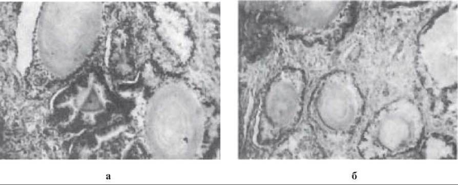
Fig. 49. Prostatic calculi.
According to O. V. Proskura (1970), sexual dysfunctions occur in 21% of cases involving prostatic calculi. Patients report exacerbated pain during sexual intercourse and defecation, as well as frequent painful pollutions.
If stone-induced Atrophy of the prostate occurs, the gland is transformed into a sac filled with calculi. Pain significantly worsens when the patient is in a sitting position.
When a concretion obliterates the urethral lumen, Acute Urinary Retention may ensue. In some instances, terminal hematuria and Urinary Incontinence are observed.
Diagnosis is established based on radiography and ultrasound findings (plain radiographs and imaging reveal calcifications in the prostatic region), as well as digital rectal examination (Palpation reveals a nodular prostate, with palpable concretions and noticeable crepitus).
Treatment. When small stones are detected in the gland accompanied by prostatitis, appropriate conservative management is prescribed (including etiotropic therapy, pathogenetic treatments, physical therapy, and health resort treatment as indicated). Therapeutic efficacy is enhanced by interventions aimed at resolving complications. It is advisable to incorporate agents that correct hemodynamic disorders and improve microcirculation, as well as antihistamines and enzyme preparations, into the comprehensive treatment regimen.
Symptomatic stones causing significant discomfort to the patient are removed surgically. Surgical intervention is indicated in cases of abscess formation and urinary retention. For calculi of the prostatic urethra, surgery to excise the anterior wall of the urethra with stone extraction is indicated.
Last update: 10/08/2026
Editorial and Educational Adaptation: This material has been compiled based on the primary/original source text. The project team performed an editorial review, corrected technical inaccuracies, structured sections, and adapted the content for an educational format.
What was processed:
- elimination of formatting defects (OCR errors, structural breaks, corrupted characters);
- editorial organization of content;
- standardization of terminology in accordance with academic sources;
- verification of factual statements against the original source text.
All mentions of the author, publication year, and origin of the primary text have been preserved in accordance with the source.