Human Anatomy, Part 1 - K. A. Dyubenko, A. K. Kolomiyytsev, Yu. B. Chaykovsky 2002
Special Section
Reproductive Systems, systemata genitalia — Female Reproductive System, systema genitale femininum
External Female Genital Organs, organa genitalia feminina externa — Pelvic Cavity — Muscles of the Perineum
The urogenital and pelvic diaphragms are formed by Muscles arranged in two layers: superficial and deep.
Muscles of the urogenital Diaphragm:
Superficial muscles (Figs. 333, 334)
- bulbospongiosus Muscle, m. bulbospongiosus;
- ischiocavernosus muscle, m. ischocavernosus;
- superficial transverse perineal muscle, m. transversus perinei superficialis.
Deep muscles (Fig. 335)
- deep transverse perineal muscle, m. transversus perinei profundus;
- urethral sphincter muscle, m. sphincter urethrae.
The bulbospongiosus muscle, m. bulbospongiosus, is a paired muscle that covers the inferolateral surface of the bulbus Penis and the corpus spongiosum, fusing with its counterpart on the opposite side to form a median raphe. The bulbospongiosus compresses the Urethra and AIDS in the evacuation of urine and semen. In females, the m. bulbospongiosus is divided into two symmetrical halves that flank and constrict the vaginal opening.
Class="center">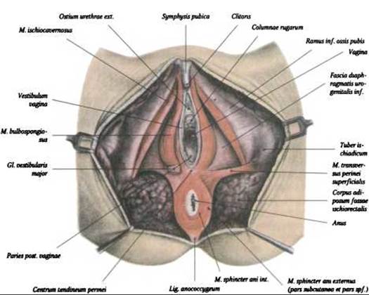
Fig. 333. SUPERFICIAL MUSCLES OF the female Perineum (after F. Kiss and J. Szentágothai)
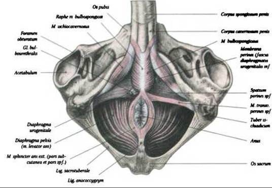
Fig. 334. Superficial muscles of the male perineum (after F. Kiss and J. Szentágothai)
The ischiocavernosus muscle, m. ischiocavernosus, originates from the ischial tuberosity on the right and left sides and inserts into the corpus cavernosum. The ischiocavernosus compresses the venous Vessels of the penis or Clitoris, thereby facilitating erection.
The superficial transverse perineal muscle, m. transversus perinei superficialis, is a small, paired muscle located at the boundary between the two diaphragms. It originates from the ischial tuberosity on both sides and extends to the perineal body, which reinforces it.
The deep transverse perineal muscle, m. transversus perinei profundus, is a flat muscle that encloses the membranous part of the urethra. It originates from the ischial tuberosities and adjacent PARTS OF THE ischial bones, extending medially to form a tendinous raphe. The deep transverse perineal muscle reinforces the urogenital diaphragm. The urethra and Vagina pass through it. In the area where the urethra pierces the muscle, some fibers of the deep perineal muscle form the urethral sphincter muscle, m. sphincter urethrae, which acts as a voluntary sphincter.

Fig. 335. Deep Muscles of the female perineum (after F. Kiss and J. Szentágothai)
Deep muscles
- levator ani muscle, m. levator ani;
- coccygeus muscle, m. coccygeus.
The levator ani muscle, m. levator ani, is a flat, triangular muscle that originates from the inferior ramus of the pubis, the Fascia of the obturator internus, and the pelvic surface of the ischial spine. One portion of its muscle fibers extends posteriorly, flanks the rectum, and fuses with the fibers of the opposite side. Another portion passes laterally to the Urinary Bladder and prostate (and in females, the vagina). These muscle fibers interlace with the musculature of the urinary bladder and vagina, extending posteriorly to insert into the coccyx and the anococcygeal ligament.
The coccygeus muscle, m. coccygeus, is located in the posterior part of the pelvic diaphragm. It originates from the ischial spine and sacrospinous ligament and inserts into the lateral margin of the sacrum and coccyx.
Muscles of the pelvic diaphragm (see Fig. 335):
Superficial muscles
- external anal sphincter, m. sphincter ani externus.
The external anal sphincter, m. sphincter ani externus, belongs to the superficial muscles of the pelvic diaphragm. It surrounds the anus, lying superficial to the m. levator ani. The muscle Functions as the external voluntary sphincter of the anus, closing it. The muscle bundles of the m. sphincter ani externus form a semi-oval shape. The superficial bundles terminate in the subcutaneous tissue, originating from the coccyx and inserting into the perineal body (central tendon of the perineum), while the deepest fibers encircle the anus and closely adhere to the m. levator ani.
Last update: 08/08/2026
Editorial and Educational Adaptation: This material has been compiled based on the primary/original source text. The project team performed an editorial review, corrected technical inaccuracies, structured sections, and adapted the content for an educational format.
What was processed:
- elimination of formatting defects (OCR errors, structural breaks, corrupted characters);
- editorial organization of content;
- standardization of terminology in accordance with academic sources;
- verification of factual statements against the original source text.
All mentions of the author, publication year, and origin of the primary text have been preserved in accordance with the source.