Human Anatomy Part 1 - K. A. Dyubenko, A. K. Kolomiytsev, Yu. B. Chaykovsky 2002
Special Part
Respiratory System, systema respiratorium [respiratory apparatus, apparatus respiratorius] - General Overview
Thoracic cavity, pleura, mediastinum - X-ray anatomy of the lungs
The posteroanterior chest radiograph clearly demonstrates the right and left pulmonary fields (Fig. 297). The apical regions of these fields project above the clavicular shadows and are referred to as the lung apices. Normally, the right pulmonary field is wider and shorter than the left. Each pulmonary field is conventionally divided into three zones: upper, middle, and lower. The Lungs of a healthy individual are air-filled, which accounts for their radiolucent (light) appearance on the radiograph. On the Medial surface of the lungs, between the anterior ends of the II and IV Ribs, a dense shadow is visible, representing the lung roots. On the right side, this shadow is crescent-shaped, whereas the shadow of the left ROOT is positioned slightly higher and is partially obscured by the cardiac silhouette. Against the Background of the pulmonary fields, a distinctive pulmonary vascular pattern formed by Arteries and Veins can be traced. This pattern exhibits typical branching, sharp contours, and a gradual tapering of vessel diameters toward the periphery. Extending from the mediastinal shadow to the lateral chest wall is the arched shadow of the Diaphragm. The dome of the diaphragm and the lateral chest wall together form an acute angle corresponding to the costodiaphragmatic recess.
Class="center">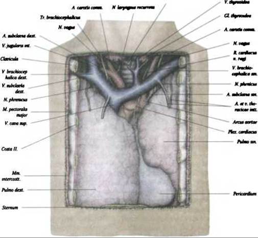
Fig. 295. Thoracic cavity, cavitas thoracis (anterior Mediastinum) (after F. Kiss)
Computed Tomography. Computed tomography provides a much clearer and more detailed Assessment of the anatomy and spatial relationships of thoracic Organs than conventional radiography. CT allows for precise measurement of vessel diameters, which is essential in the Diagnosis of cardiovascular diseases. CT scans of the chest clearly delineate the pulmonary parenchyma, mediastinal organs, as well as the bones and Cartilage of The thoracic cage. The resulting image varies depending on the level of the cross-section.
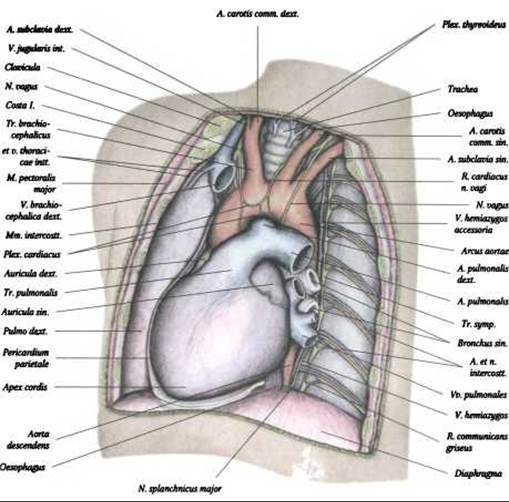
Fig. 296. Thoracic cavity, cavitas thoracis (middle and posterior mediastinum) (after F. Kiss)
At the level of the sternoclavicular joint, anterior to the Trachea, the brachiocephalic veins are visible. Situated between the left brachiocephalic vein and the anterolateral surface of the trachea are three major arterial trunks: the brachiocephalic trunk, the left common carotid artery, and the left Subclavian Artery. The Esophagus can be seen lying between the posterior wall of the trachea and the vertebral bodies. Small Lymph Nodes may be identified within the paratracheal adipose tissue. Due to the predominantly vertical and oblique orientation of the vessels, the pulmonary vasculature appears on scans as cross-sections and short segments branching into finer vessels.
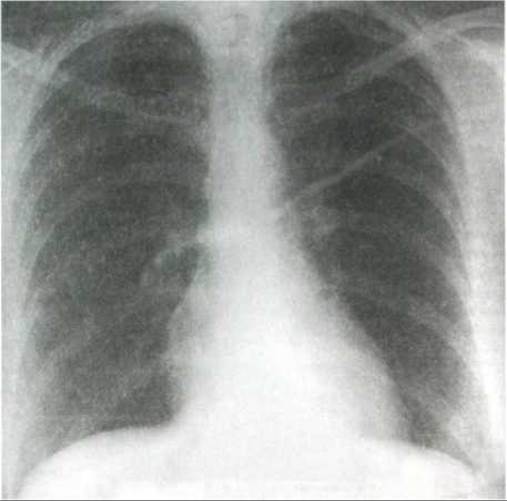
Fig. 297. Posteroanterior chest radiograph (after A. V. Makarov)
At the level of the tracheal bifurcation, posterior and slightly to the left of it, lies the esophagus. In slices taken slightly more inferiorly, the descending part of the aortic arch abuts the posterior wall of the left main bronchus. On the right, at the same level, the cross-section of the azygos vein is visualized with a diameter of approximately 0.6 cm.
Last update: 08/08/2026
Editorial and Educational Adaptation: This material has been compiled based on the primary/original source text. The project team performed an editorial review, corrected technical inaccuracies, structured sections, and adapted the content for an educational format.
What was processed:
- elimination of formatting defects (OCR errors, structural breaks, corrupted characters);
- editorial organization of content;
- standardization of terminology in accordance with academic sources;
- verification of factual statements against the original source text.
All mentions of the author, publication year, and origin of the primary text have been preserved in accordance with the source.