Human Anatomy, Part 1 - K. A. Dyubenko, A. K. Kolomiytsev, Yu. B. Chaykovsky 2002
Special Part
Respiratory System, systema respiratorium [respiratory apparatus, apparatus respiratorius] - General Overview
Bronchi
The right and left main Bronchi originate from the Trachea: bronchus principalis dexter et sinister (see Fig. 287). The right bronchus is shorter and wider than the left. The length of the right bronchus is 1.5–2 cm, with a diameter of 1.5–2.5 cm, while for the left, these values are 4–6 cm and 1–2 cm, respectively. The angle between the trachea and the right bronchus is 150–160∘, and between the trachea and the left bronchus, 130–140∘. Due to the fact that the right bronchus is positioned more vertically and has a larger diameter, Foreign bodies in the Airways more frequently enter the right lung through it.
The azygos vein arches over the superior surface of the right bronchus, and tracheobronchial Lymph Nodes lie adjacent to this same surface. The posterior surface of the right bronchus is related to the right Vagus nerve and the right posterior bronchial artery; the anterior surface to the ascending aorta, the right anterior bronchial artery, and the Pericardium; and the inferior surface to the bifurcation lymph nodes. The aortic arch is adjacent to the superior surface of the left bronchus, the descending aorta, the left vagus nerve, and the Esophagus to the posterior surface, the left anterior bronchial artery and tracheobronchial lymph nodes to the anterior surface, and the bifurcation lymph nodes to the inferior surface.
The wall Structure OF THE main bronchi is similar to that of the trachea. Its framework consists of cartilaginous rings, of which there are 6–8 in the right bronchus and 9–12 in the left. The mucous membrane is lined with pseudostratified ciliated epithelium.
During bronchoscopy, that is, when examining the internal surface of the bronchi using a bronchoscope, the mucous membrane has a grayish-pink color. The carina tracheae, the projection between the walls of the main bronchi at their origin, is clearly visible.
In clinical (pulmonological) practice, bronchography of the Lungs is performed in anteroposterior and lateral projections, allowing for the visualization of the main, lobar, and segmental bronchi (Figs. 288, 289).
In newborns, the bronchi are wide. During the first year of life, they grow very rapidly, and then relatively slowly until the age of 9–10. By age 13, the length of the bronchi doubles.
The Blood supply to the bronchi is provided by the bronchial Arteries, and venous blood drainage is carried out by the corresponding Veins.
Lymph from the bronchi drains into the bronchopulmonary, tracheobronchial, and bifurcation lymph nodes.
The Innervation of the bronchi is provided by the sympathetic and parasympathetic Branches of the bronchial plexus.
Class="center">
Fig. 288. Bronchogram of the left lung in lateral projection (according to A. V. Makarov). The main, lobar, and segmental bronchi are contrasted (normal)
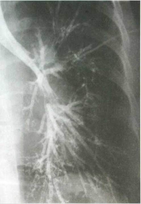
Fig. 289. Bronchogram of the left lung in anteroposterior projection (according to A. V. Makarov). The main, lobar (superior and inferior), and segmental bronchi are contrasted (normal)
Last update: 08/08/2026
Editorial and Educational Adaptation: This material has been compiled based on the primary/original source text. The project team performed an editorial review, corrected technical inaccuracies, structured sections, and adapted the content for an educational format.
What was processed:
- elimination of formatting defects (OCR errors, structural breaks, corrupted characters);
- editorial organization of content;
- standardization of terminology in accordance with academic sources;
- verification of factual statements against the original source text.
All mentions of the author, publication year, and origin of the primary text have been preserved in accordance with the source.