MEDICAL BIOLOGY, ANATOMY, HUMAN PHYSIOLOGY AND PATHOLOGY - Ya.I.Fedoniuk 2010
BIOLOGY
CHAPTER 1. BIOLOGICAL FOUNDATIONS OF HUMAN VITAL ACTIVITY
1.4. ONTOGENETIC LEVEL OF LIFE ORGANIZATION
1.4.2. Fundamentals of human genetics
Gene Mutations
These are Changes in the Chemical Structure of a gene (DNA). If the changes affect only a single nucleotide in DNA, such mutations are called point mutations. Gene mutations are not visible under a Microscope and are detected by the appearance of an altered trait in the offspring controlled by the mutant gene. Mutations of this type occur most frequently. They give rise to new alleles, increase the gene pool of populations, and are significant for evolution.
The MAIN TYPES OF gene mutations include substitutions, insertions, deletions, and duplications of nucleotide pairs. In all cases, they alter the DNA nucleotide sequence. These changes are transcribed into an mRNA molecule and often lead to The formation of an altered polypeptide. The most typical gene mutations are base substitutions and reading frameshift mutations. The latter are associated with the loss or insertion of one or more NUCLEOTIDES. Mutations in which a purine base is replaced by a purine (A<->G) or a pyrimidine base by a pyrimidine (T<->C) are called transitions. When a purine base is replaced by a pyrimidine and vice versa, the mutations are called transversions (A<->T, A<->C, G<->C, and G<->T). Base substitution mutations lead to the appearance of missense and nonsense mutations. A missense mutation is a nucleotide substitution in the coding system of a gene that can cause the replacement of an amino acid in a protein molecule. A nonsense mutation is a nucleotide substitution in the coding part of a gene that leads to the formation of a nonsense (stop) codon. There are mutations where a nucleotide substitution in the coding part of a gene does not alter the meaning of METABOLISM/28.html">The Genetic Code due to its redundancy. However, in the case of Sickle Cell anemia, this is not true (Fig. 1.80).
Class="center">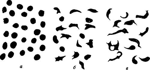
Fig. 1.80. Sickle cell anemia
Sickle cell congenital anemia. It is called sickle cell (SCA) because Blood smears reveal erythrocytes in the shape of a sickle. It was later established that the disease is a gene mutation and is inherited in an autosomal recessive manner. In this condition, erythrocytes contain abnormal Hemoglobin S. The protein part of the adult human hemoglobin molecule consists of four polypeptide chains (two alpha-chains and two beta-chains) in patients with sickle cell anemia. Each alpha-chain contains 141 Amino Acids, and each beta-chain contains 146. These chains are encoded by different genes. Abnormal hemoglobin Hbs differs from normal HbA by the substitution of a single amino acid (in the beta-chain, glutamic acid at the sixth position is replaced by valine — Glu<->Val). The whole molecule consists of 574 amino acids. The difference is only in Two amino acids (one in each of the two beta-chains). This single difference between HbA and Hbs is explained by the replacement of a single base in the corresponding gene triplet. Glutamic acid is coded by two codons — GAA and GAG, and valine by four — GUU, GUC, GUA, and GUG, meaning a mutation occurred involving the substitution of the second Base of the codon (A<->U). This seemingly very small difference causes fatal consequences. Erythrocytes with abnormal hemoglobin, instead of their normal biconcave disc shape, acquire a sickle shape, become fragile, break down rapidly, and are incapable of transporting oxygen. In homozygous individuals HbsHbs, severe SCA develops, and they die in early childhood. Heterozygotes HbAHbs are clinically healthy, but under conditions of oxygen deficiency (high altitudes, diving, aviation), symptoms of anemia manifest in them. This is because a significant portion of the heterozygotes' erythrocytes contains abnormal hemoglobin (Fig. 1.80).
Gene mutations also cause hemophilia, color blindness, albinism, phenylketonuria, alkaptonuria, and galactosemia. These diseases are called genetic or molecular because they are caused by changes in the gene structure (DNA molecule).
Last update: 08/08/2026
Editorial and Educational Adaptation: This material has been compiled based on the primary/original source text. The project team performed an editorial review, corrected technical inaccuracies, structured sections, and adapted the content for an educational format.
What was processed:
- elimination of formatting defects (OCR errors, structural breaks, corrupted characters);
- editorial organization of content;
- standardization of terminology in accordance with academic sources;
- verification of factual statements against the original source text.
All mentions of the author, publication year, and origin of the primary text have been preserved in accordance with the source.