BIOLOGY Volume 3 - A Textbook of General Biology - 2004
21. REPRODUCTION
21.8. Human reproduction
21.8.5. Implantation
As it travels down the fallopian tube toward the Uterus, the zygote undergoes a series of successive nuclear and cellular divisions, resulting in a small ball of Cells; this process is known as Cleavage. During cleavage, the cells divide without increasing in overall size, as they remain confined within the zona pellucida. At this stage, the cells become progressively smaller with each division, while their nuclei divide mitotically. The resulting cells are called blastomeres financières. After A number of divisions, the blastomeres form a hollow sphere with a central cavity known as the blastocoel. The outer layer of cells, called the trophoblast, differentiates in one region to form the inner Cell mass. This stage of development, reached 4–5 days after Fertilization, is called the blastocyst. The Structure of the blastocyst is shown in Fig. 21.47.
Class="center">
Fig. 21.47. Simplified diagram of the structure of a human blastocyst four days after fertilization.
Upon entering the uterus, the blastocyst remains in the uterine cavity for about two days; during this time, the zona pellucida gradually disintegrates, allowing the trophoblast cells (from the Greek trophé, meaning nourishment) to come into direct contact with the endometrial cells. Nourished by the endometrium, the trophoblast cells proliferate, and by the 6th to 9th day following fertilization, the blastocyst becomes fully embedded in the endometrium. This process is called implantation.
The trophoblast cells differentiate into an inner and an outer layer. The outer layer, termed the chorion, gives rise to trophoblastic villi (chorionic villi)—finger-like projections that invade the endometrium (Fig. 21.48). The Regions of the endometrium between these villi form interconnecting spaces called lacunae, giving this area of the endometrium a spongy appearance. Under the action of hydrolytic Enzymes secreted by the trophoblast, the maternal arterial and venous vessels in the endometrium rupture, and the escaping Blood fills the lacunae. In the Cytology/cytology/16.html">Early stages of blastocyst development, the exchange of nutrients, oxygen, and Metabolic waste products between the blastocyst cells and maternal blood occurs across the chorionic villi of the trophoblast. At later Selection/3.html">Stages of development, this function is taken over by the Placenta.
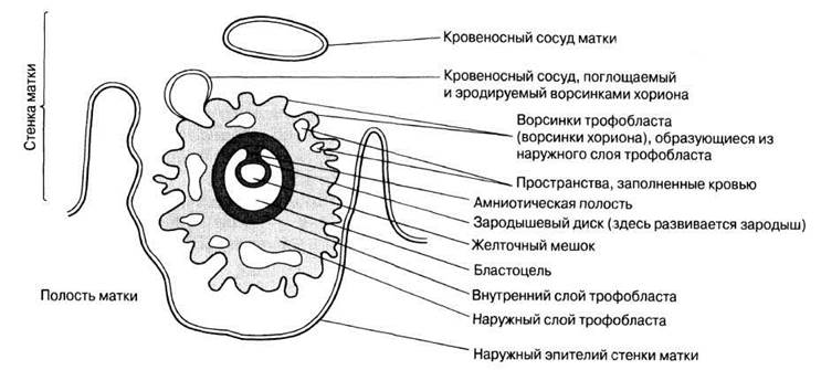
Fig. 21.48. Human blastocyst shortly after implantation into the uterine endometrium (simplified diagram). Enzymes produced by the outer layer of the trophoblast (the chorion) break down the endometrial Blood Vessels, creating blood-filled spaces that supply the blastocyst with nutrients and remove metabolic waste products.
Last update: 06/08/2026
Editorial and Educational Adaptation: This material has been compiled based on the primary/original source text. The project team performed an editorial review, corrected technical inaccuracies, structured sections, and adapted the content for an educational format.
What was processed:
- elimination of formatting defects (OCR errors, structural breaks, corrupted characters);
- editorial organization of content;
- standardization of terminology in accordance with academic sources;
- verification of factual statements against the original source text.
All mentions of the author, publication year, and origin of the primary text have been preserved in accordance with the source.