BIOLOGY Volume 2 - A Guide to General Biology - 2004
15. HEALTH AND DISEASE
15.3. Infectious Diseases
15.3.2. Tuberculosis
Tuberculosis is one of the earliest known fatal human diseases. Evidence of it has been found in the skeletons of people who lived as long ago as the late Stone Age (Neolithic period). Fig. 15.5 shows tuberculosis mortality rates in England and Wales from 1836 to 1970. In the 19th century, it was one of the leading causes of death, and in England and Wales the primary one, claiming the lives of one in five people. By 1990, childhood mortality from tuberculosis in developed countries had dropped to less than 1 case per 1 million, yet worldwide at least 30 million people continue to suffer from the disease, 95% of them in developing countries. Annually, the disease claims up to 3 million lives, and approximately every third person on the planet is an asymptomatic carrier of the pathogen.
Class="center">
Fig. 15.5. Dynamics of mortality from Pulmonary Tuberculosis in England and Wales from 1838 to 1970.
In the early 1990s, the WHO declared tuberculosis a global threat as the number of cases in developed countries, including Britain, began to rise again in parallel with increasing drug resistance of the pathogen. In 1992, 5,802 newly diagnosed cases were registered in England, and worldwide the number of new cases reaches 8 million annually. In 1993, Dr. Kochi, director of the WHO tuberculosis programme, stated: "Tuberculosis has slipped out of control in many regions. This disease, whose Prevention and Treatment Methods have long been known, is not receiving adequate attention, and no country is safe from it." The global distribution of tuberculosis mortality and incidence is shown in Fig. 15.6.

Fig. 15.6. New cases of tuberculosis and mortality from it in certain Regions of the world in the late 1980s. (From World Health Organisation, 1989/90.)
The disease is caused by the bacterium Mycobacterium tuberculosis, belonging to the actinomycete group (like Fungi, they form hyphae). It was discovered by Robert Koch in 1882, which is why the microorganism is historically known as "Koch's bacillus". It is also sometimes referred to as the tubercle bacillus (a bacillus being any rod-shaped bacterium), though "tuberculosis mycobacterium" is more accurate. This pathogen can affect many Organs, but the most common form of the disease is pulmonary tuberculosis. Two strains hazardous to humans are known: the human strain and the bovine strain (sometimes considered a distinct species, M. bovis). The latter primarily affects cattle and can find its way into milk. It is highly resilient and retains its virulence in dairy products for a long time. In the past, alongside livestock losses, the bovine strain contributed significantly to human morbidity and mortality, particularly among children. However, all milk in the UK is now sourced from cows that have undergone the tuberculin test, which certifies the absence of the pathogen. Furthermore, before going on sale, milk is sanitised by pasteurisation, sterilisation, or ultra-high-Temperature (UHT) Processing. These methods destroy at least 99% of all Bacteria, including pathogens. Consequently, bovine tuberculosis no longer poses a threat to humans in developed countries.
Transmission Routes and Symptomatology
Pulmonary tuberculosis is airborne, meaning a person becomes infected by inhaling air containing droplets of saliva from an infected individual. The tuberculosis mycobacterium is considerably less virulent than common cold pathogens, and prolonged contact between individuals is required for transmission. It withstands desiccation, allowing it to accumulate and persist for long periods in indoor air and dust. For this reason, the disease primarily affects those living in overcrowded conditions. Typical settings for the spread of tuberculosis include refugee and prisoner-of-war camps, homeless shelters, and prison Cells. In such environments, susceptibility is further heightened by malnutrition and the presence of other infections, which collectively weaken the body's immune system.
Tuberculosis can affect almost any organ, but the Lungs are affected far more frequently than any others. In the past, the pulmonary form of tuberculosis was known as "consumption" because the patient visibly "wasted away", losing weight and strength. The outcome of the disease depends on a variety of factors, including the person's age, nutritional status (usually linked to their social standing), and the state of their immune system. Immunity to tuberculosis develops As a result of a mild infection or vaccination (see below).
The disease often manifests with non-specific symptoms such as loss of weight, lack of appetite, and heavy sweating. In its early stages, tuberculosis is generally asymptomatic and may be detected incidentally during a chest X-ray or fluorography (Fig. 15.7). The disease begins with foci of inflammation in the lungs, leading to The formation of nodular scars or tissue necrosis, which results in The Development of cavities. These expand and multiply, ultimately leading to the characteristic clinical picture: a cough, hemoptysis, chest pain, shortness of breath, accompanied by fever, sweating, and loss of appetite and weight.
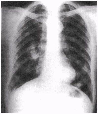
Fig. 15.7. Chest X-ray of a patient with pulmonary tuberculosis. A normal lung would be uniformly dark. The light patches against the dark Background correspond to infected areas.
Treatment and Prevention
An effective drug against tuberculosis—the antibiotic streptomycin—did not appear until 1947. Mass vaccination began in the UK in 1954. The decline in incidence observed even earlier was primarily associated with improving living conditions, especially housing. Vaccination accelerated this decline (Fig. 15.5), and by 1970 only 1,526 people died from tuberculosis in Britain.
VACCINATION. The world owes the creation of an effective tuberculosis vaccine to the French scientists Albert Calmette and Camille Guérin. Named after them, the preparation is called the "Bacille Calmette-Guérin", or BCG for short. As early as 1921, they obtained attenuated (weakened, low-virulence) strains of the tuberculosis mycobacterium suitable for active immunization. Before vaccination, It is important to check whether the person currently has the disease or has already acquired immunity. To do this, a special instrument (a ring of six short needles) is used to introduce the protein tuberculin, extracted from killed mycobacteria, into the Skin1. If the individual has no Antibodies against it, no characteristic reaction is observed (a negative tuberculin test); if antibodies are present, the skin reddens and swells at the injection site (a positive tuberculin test). This indicates that immunity has developed naturally and vaccination is unnecessary.
Long-term observations of 50,000 initially healthy children showed that the prevalence of tuberculosis is 1.91 per 1,000 in the unvaccinated group, compared to 0.4 per 1,000 in the vaccinated group, with artificial immunity lasting over 10 years. Vaccination is currently administered to children aged 12–14 (Table 15.2). Tuberculin tests at this age yield positive results in approximately 10% of cases. These children are scheduled for standard fluorography to check for active tuberculosis, which is very rarely detected.
Antibiotics. An effective cure for tuberculosis was unknown until 1943, when the antibiotic streptomycin was discovered. Following this, incidence rates began to drop noticeably (Fig. 15.5). This trend continued until the mid-1980s, facilitated by the Introduction of Other Antibiotics, notably rifampicin and isoniazid. By this time in Western countries, over 80% of all active tuberculosis patients were people over the age of 60.
Rising Incidence
After 1980, the disease began spreading to increasingly younger age groups, reaching individuals aged 25–30. From 1980 to 1986, five separate studies in the US demonstrated a correlation between the increase in the homeless population and tuberculosis incidence among young people. By 1985, it became clear that in many cases the disease was caused by new mutant strains of mycobacteria resistant to drugs. While in 1986 strains resistant to isoniazid and rifampicin caused 0.5% of tuberculosis cases, by 1991 this figure had risen to 3%, and by 1994 to 6.9%. The main factors contributing to this were overly short treatment courses that failed to kill all mycobacteria, and patient non-compliance with doctors' instructions. A full course of treatment lasts 6–8 months and requires taking multiple tablets daily. This involves a combination of three to four antibiotics, which reduces the likelihood of strains surviving that are resistant to any single one of them. The challenge lies in the fact that patients begin to feel better after a few weeks and arbitrarily stop treatment. Combating this phenomenon is difficult not only in developing countries but also in the US, since the majority of tuberculosis patients are homeless. As a result, the disease is once again assuming epidemic proportions in some regions.
From the very beginning of the AIDS epidemic, a correlation began to emerge between HIV carriage and tuberculosis (Section 15.3.4). However, despite WHO warnings regarding The Link Between HIV and this disease, authorities in the US and Western Europe long continued to view the elevated risk of tuberculosis in HIV-infected individuals as exclusively a Third World problem. Tuberculosis did indeed begin to spread rapidly in Africa, and HIV patients responded poorly to cheap anti-tuberculosis drugs such as thiacetazone and streptomycin. By 1990, public health officials in several African countries declared themselves powerless to cope with the epidemic.
New drug-resistant strains of mycobacteria are spreading rapidly and show a clear link to HIV infection. AIDS patients are highly susceptible to tuberculosis, with mortality rates reaching 90–100% among them. The rise in tuberculosis incidence in several European countries is reflected in Table 15.7, showing a direct correlation with the spread of resistant pathogen strains and HIV infection.
Table 15.7. Increase in tuberculosis incidence in developed countries according to WHO data (World Health Organisation, Press Release June 17, 1992)
Country |
Period |
Increase, % |
Switzerland |
1986-1990 |
33 |
Denmark |
1984-1990 |
31 |
Italy |
1988-1990 |
28 |
Norway |
1988-1991 |
21 |
Ireland |
1988-1990 |
18 |
Austria |
1988-1990 |
17 |
Finland |
1988-1990 |
17 |
USA |
1986-1991 |
12 |
Netherlands |
1987-1990 |
9,5 |
Sweden |
1988-1990 |
4,6 |
United Kingdom |
1987-1990 |
2,0 |
France, Germany, Belgium |
1987-1991 |
0 |
A 1996 report on tuberculosis incidence in Edinburgh for the period 1988–1992 demonstrates the following:
1) among individuals over 65 years of age, the number of cases increased by 4.1%;
2) among younger individuals, this increase was 12.6%.
Tuberculosis in older adults is quite frequently the result of the reactivation of a childhood or youth infection. In both the elderly and the young, the rise in incidence is partly driven by the increasing resistance of tubercle bacilli to antibiotics.
The number of tuberculosis cases is also growing in connection with immigration. For example, in areas of the UK densely populated by recent immigrants from former colonies, the increase in incidence is 25% higher than in areas with a predominantly native population.
To improve treatment outcomes for tuberculosis, the WHO has introduced a directly observed therapy (DOT) strategy. A healthcare worker personally gives the pills to the patient and ensures they swallow them. This form of treatment, lasting 6–8 months, leads to recovery in over 85% of cases.
1 In our country, the Mantoux test is also widely used, in which tuberculin is injected intradermally using a standard syringe. — Transl. note.
Last update: 06/08/2026
Editorial and Educational Adaptation: This material has been compiled based on the primary/original source text. The project team performed an editorial review, corrected technical inaccuracies, structured sections, and adapted the content for an educational format.
What was processed:
- elimination of formatting defects (OCR errors, structural breaks, corrupted characters);
- editorial organization of content;
- standardization of terminology in accordance with academic sources;
- verification of factual statements against the original source text.
All mentions of the author, publication year, and origin of the primary text have been preserved in accordance with the source.