BIOLOGY Volume 1 - A Guide to General Biology - 2004
9. UTILIZATION OF ENERGY
9.5. Gas exchange in mammals
9.5.1. Structure of the respiratory system
The respiratory surface in mammals is formed by numerous air-filled sacs known as alveoli. The alveoli are located inside the paired Lungs, which are situated in the thoracic cavity close to The Heart and communicate with the outside air via a series of air passages (Fig. 9.19). Air enters the lungs through these tubes. The heart and lungs are surrounded and protected by twelve pairs of bony Ribs. Attached to the ribs are the intercostal Muscles, and the thoracic cavity is separated from the Abdominal cavity by a broad, dome-shaped Diaphragm. These muscles and the diaphragm also play an essential role in the ventilation mechanism, which is discussed in section 9.5.4.
Class="center">
Fig. 9.19. Human Trachea and lungs.
Air enters the body through the two nostrils, each of which is lined with a fringe of hairs that filter out foreign particles. As the air passes through the nasal passages, it is warmed and moistened. At the same time, the animal samples airborne odors. The air then passes through the Larynx into the trachea. The trachea is a tube that lies immediately ventral to the Esophagus and extends down into the chest cavity. Its walls are reinforced with C-shaped cartilaginous rings, which keep the airway permanently open. The open ends of the C-shaped rings face the esophagus (Fig. 9.19); this prevents the trachea from collapsing during inhalation. Such a Cartilage ring can be seen in a transverse section of the trachea (Fig. 9.20, A).
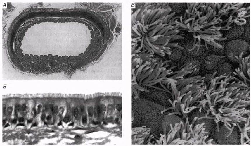
Fig. 9.20. A. Transverse section of the trachea (light micrograph, low power). B. Ciliated epithelium with goblet Cells (light micrograph, high power). C. Surface of the ciliated epithelium (scanning electron micrograph).
At its lower end, the trachea divides into two Bronchi. Within the lungs, each bronchus branches repeatedly into smaller tubes called bronchioles. These in turn subdivide into progressively finer tubules that terminate in alveolar ducts leading into alveolar sacs. Each sac contains a cluster of individual alveoli that open into it. Figure 9.21 illustrates this pathway of airflow along with the histological features of its various regions.

Fig. 9.21. Histological CHARACTERISTICS OF THE respiratory pathway.
The walls of most of the respiratory tract are lined with ciliated epithelium. Interspersed within this epithelium are goblet cells that secrete mucus (Figs. 9.20 and 6.16). The mucus traps any particles that manage to bypass the nasal hairs, such as dust and Bacteria. The beating of the cilia moves these mucus-trapped particles up to the back of the Pharynx, where they are swallowed (thus preventing them from entering the lower Airways). Note that foreign particles here are trapped by mucus rather than by the cilia, whereas in the Nasal cavity they are filtered out directly by hairs. Mucus also performs another function: it humidifies the incoming inspired air.
Structure OF THE alveoli
The walls of the alveoli serve as the primary site for gas exchange (Fig. 9.22). Human lungs contain up to 700 million alveoli, providing a total surface area of 70–90 $m^2$. The thickness of the alveolar wall is only about 0.0001 mm (0.1 µm). The outer surface of the alveolar wall is enveloped in a dense network of Blood capillaries; all of these originate from the pulmonary artery and eventually converge to form the pulmonary vein (Figs. 9.23 and 9.24). Each alveolus is lined with a moist squamous epithelium. Its cells are extremely flattened (Fig. 6.14), which further minimizes the diffusion distance for gases (Fig. 9.24). The alveolar wall also contains Collagen and elastic fibers, giving it flexibility and allowing the alveoli to change volume during inspiration and expiration.
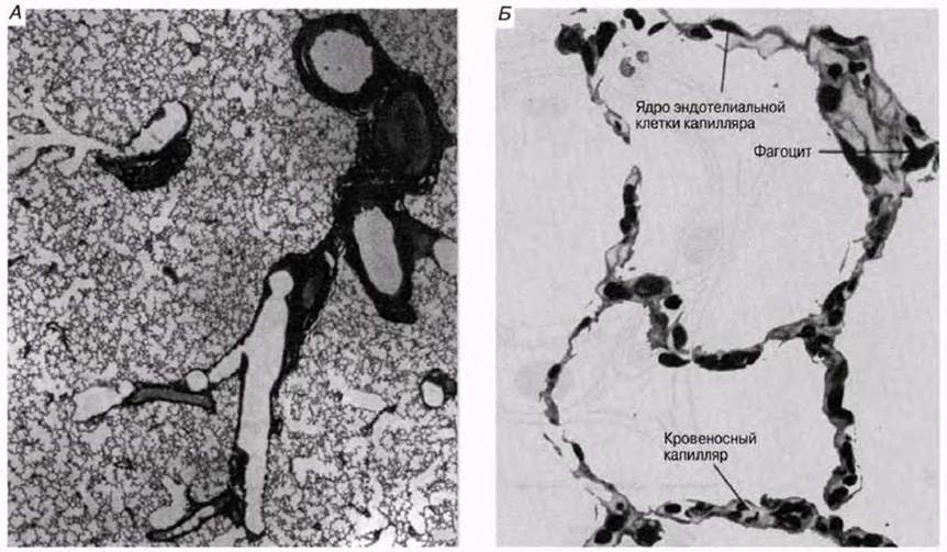
Fig. 9.22. A. Human lung tissue under a Light Microscope at low magnification. B. Alveoli at high magnification.
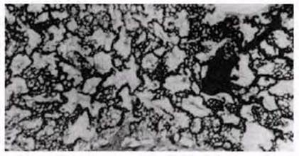
Fig. 9.23. Human lung injected with a specialized contrast medium to reveal the blood vessel network.

Fig. 9.24. A. Cross-section of an alveolus. Parts of five adjacent alveoli and various interalveolar structures are also shown. B. The relationship between an alveolus and a capillary.
Specialized cells within the alveolar wall secrete a detergent-like substance known as surfactant onto its inner surface. This substance reduces the surface tension of the moisture layer lining the alveolar epithelium, thereby decreasing the muscular effort required to expand the lungs during breathing. Surfactant also accelerates The transport of oxygen and CO2 across this fluid layer. In addition, it helps destroy bacteria that manage to reach the alveoli. In healthy lungs, surfactant is continuously secreted and reabsorbed. In human fetuses, it first appears around the 23rd week of gestation. This is a key reason why a fetus prior to 24 weeks is considered non-viable. It also dictates the legal threshold in the UK before which elective premature labor induction is prohibited. It is believed that infants born prematurely may lack sufficient surfactant. Consequently, this leads to respiratory distress syndrome, one of the leading causes of death in premature newborns. Without surfactant, the surface tension of the fluid within the alveoli is 10 times higher than normal, causing the alveoli to collapse after every exhalation, which demands considerably more effort to reinflate them during the next breath.
Last update: 06/08/2026
Editorial and Educational Adaptation: This material has been compiled based on the primary/original source text. The project team performed an editorial review, corrected technical inaccuracies, structured sections, and adapted the content for an educational format.
What was processed:
- elimination of formatting defects (OCR errors, structural breaks, corrupted characters);
- editorial organization of content;
- standardization of terminology in accordance with academic sources;
- verification of factual statements against the original source text.
All mentions of the author, publication year, and origin of the primary text have been preserved in accordance with the source.