Protein Structure and Function. Application of Bioinformatics Methods - John Rigden 2014
Prediction of Protein Function Based on Theoretical Models
Practical Application
Structure Modeling of Alternatively Spliced Isoforms
Many, if not most, eukaryotic genes undergo Alternative Splicing, which dramatically increases The Diversity of mature molecules. It is often difficult to predict from an alternative transcript sequence whether its function will be preserved or altered. Structural modeling, where feasible, can shed light On the Relationship between Structure and function in alternative transcripts of the same Gene.
In one of the pioneering studies in this field (Fumham et al., 2004), covering models of 14 Proteins and 40 splicing variants, it was demonstrated that exon loss more frequently involves the deletion of entire structural units rather than smaller fragments. The authors showed that, according to model reliability assessment programs, modeling deletions yields more reliable results than modeling insertions. For four proteins of biomedical relevance, the authors were able to establish a correlation between the known Functional Properties of alternative variants and their structural models. Later, it was shown (Wang et al., 2005) that splicing events typically occur in loop regions on the protein surface rather than within elements of ordered Secondary structure. Generally, a given gene has only one or two splicing sites, which in 60% of cases span no more than 50 residues. These findings suggested that splicing usually occurs at sites and in a manner that preserves the protein's tertiary structure as much as possible. This is consistent with the fact that most alternative isoforms exhibit folding properties similar to the original, and therefore retain a similar function. However, a subsequent structural analysis of several transcripts revealed (Tress et al. 2007) that many isoforms should possess dramatically different structures compared to the already known structures of other isoforms. For 49 out of 85 transcripts aligned with homologous structures, the authors concluded that the isoforms and principal transcripts must form substantially different structures. An example from Tress et al. (2007) (Fig. 12.6) illustrates the interleukin-4 isoform lacking exon 2. The structural fragment encoded by this exon is part of the protein core and participates in The formation of a disulfide bond, suggesting that the three-dimensional STRUCTURE OF THE isoform will differ significantly from the known structure of the full-length protein. We still do not have a complete picture of how structural changes—whether large or small—induced by alternative splicing affect protein function. For instance, experimental data illustrating Functional differences between alternative isoforms could be found for only 4 out of 214 loci (Tress et al., 2007).
Class="center">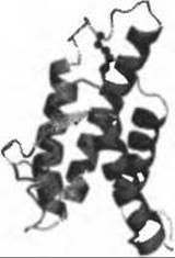
Fig. 12.6. (For the color version of this figure, see the insert.) Structure of interleukin 4 showing the fragment encoded by exon 2. The experimental structure (PDB code 1ilt) is shown in ribbon representation, with the fragment encoded by exon 2 highlighted in magenta. Disulfide Bonds are shown in stick representation; the bond within the exon 2-encoded fragment is shown in ball-and-stick representation
Last update: 06/08/2026
Editorial and Educational Adaptation: This material has been compiled based on the primary/original source text. The project team performed an editorial review, corrected technical inaccuracies, structured sections, and adapted the content for an educational format.
What was processed:
- elimination of formatting defects (OCR errors, structural breaks, corrupted characters);
- editorial organization of content;
- standardization of terminology in accordance with academic sources;
- verification of factual statements against the original source text.
All mentions of the author, publication year, and origin of the primary text have been preserved in accordance with the source.