BIOCHEMISTRY - L. Stryer - 1984
VOLUME 2
PART II GENERATION AND STORAGE OF METABOLIC ENERGY
CHAPTER 18. AMINO ACID DEGRADATION AND THE UREA CYCLE
18.12. Cobalamin (Vitamin B12)-Containing Enzymes Catalyze Rearrangement and Methylation Processes
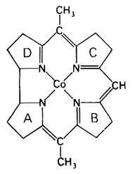
Cobalamin (vitamin B12) has attracted great interest among biochemists and physicians ever since George Minot and William Murphy discovered in 1926 that pernicious anemia could be treated by feeding large amounts of Liver to patients. Cobalamin was obtained in purified form and crystallized in 1948. Its complex three-dimensional Structure was elucidated by Dorothy Hodgkin in 1956. The core of cobalamin consists of a corrin ring with a centrally located cobalt atom (Fig. 18.12). The corrin ring, like porphyrin, contains four pyrrole rings. Two of them (rings A and D) are directly linked to each other, while the others are connected by methylene bridges, as is the case in Porphyrins. The substituents on the pyrrole rings are methyl, propionamide, and acetamide groups.
Class="center">Fig. 18.12. The corrin core of cobalamin. Substituents on the pyrroles and two other cobalt ligands are not shown in this diagram
The cobalt atom is bonded to four nitrogen atoms of the pyrrole rings. The fifth substituent (in Fig. 18.13, located below the corrin plane) is a dimethylbenzimidazole derivative containing ribose-3-phosphate and aminoisopropanol. One of the nitrogen atoms of dimethylbenzimidazole is bonded to cobalt. The amino group of aminoisopropanol is linked by an amide bond to the side chain of ring D. The sixth substituent at the cobalt atom (located above the corrin plane in Fig. 18.13) can be CN-, -CH3, OH-, or deoxyadenosyl. The presence of a cyanide ion in the sixth coordination position of cyanocobalamin, the most common commercially produced form of the vitamin, is a result of its isolation Procedure. In vivo, cyanide is not bound to cobalamin.
Fig. 18.13. Structure of 5'-deoxyadenosylcobalamin
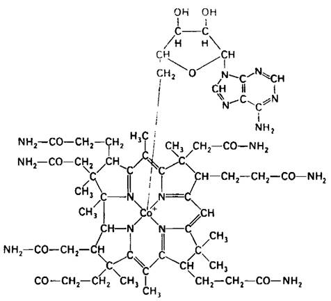
The cobalt atom in cobalamin can exist in the +1, +2, or +3 oxidation state. In hydroxocobalamin (in which the sixth coordination position is occupied by OH-), the cobalt atom is in the +3 oxidation state. This form, B12h (Co3+), is reduced to the divalent state, called B12r (Co2+), by the action of flavoprotein reductase. B12r is reduced by a second flavoprotein reductase to B12s (Co+). NADH serves as the reducing agent in both reactions. The B12s form serves as a substrate for the final enzymatic reaction leading to The formation of the active coenzyme: the 5'-deoxyadenosyl group is transferred from ATP to vitamin B12s (Co+) to form 5'-deoxyadenosylcobalamin, which is the coenzyme for methylmalonyl-CoA mutase (Fig. 18.13).
B12s (Co3+) → B12r (Co2+) → B12s (Co+) → 5'-deoxyadenosylcobalamin.
Fig. 18.14. The 5'-carbon atom of 5'-deoxyadenosine is coordinated with the cobalt atom of 5'-deoxyadenosylcobalamin. This is the only known example of a carbon-metal bond in a biomolecule
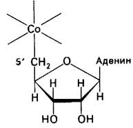
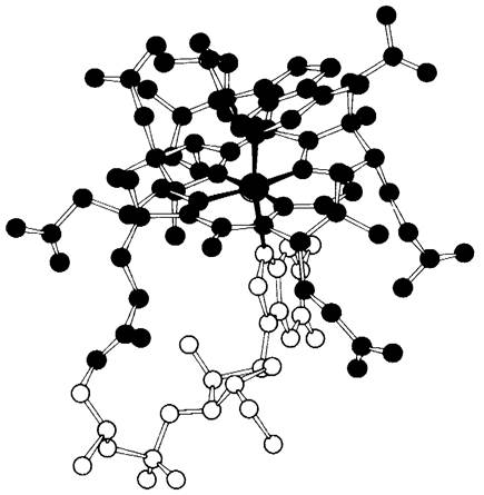
Fig. 18.15. Model of 5'-deoxyadenosylcobalamin. The cobalt atom is shown in green, the corrin component in red, the deoxyadenosyl component in blue, and the benzimidazole component in yellow
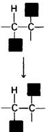
Cobalamin Enzymes catalyze Two Types of reactions: 1) rearrangement reactions, such as The conversion of L-methylmalonyl-CoA to succinyl-CoA, and 2) methylation reactions, such as the synthesis of Methionine (Section 21.7). A rearrangement represents the mutual exchange of two groups attached to adjacent carbon atoms (Fig. 18.16). A hydrogen atom migrates from one carbon atom to the next, while the R group simultaneously moves in the opposite direction. These intramolecular rearrangements involve the Cleavage of the carbon-cobalt bond in 5'-deoxyadenosylcobalamin. The carbon atom of the substrate that donates its hydrogen atom becomes temporarily bonded to cobalt.
Fig. 18.16. Rearrangement reaction catalyzed by cobalamin enzymes. The R group can be an amino group, a hydroxyl group, or any carbon-containing substituent
18.13. Impaired Cobalamin Absorption in Pernicious Anemia
Cobalamin absorption is carried out by a specialized transport system. The Stomach secretes a glycoprotein called intrinsic factor, which binds cobalamin in the intestinal lumen. The resulting complex binds to a specific receptor on the inner surface of the ileum. The cobalamin-intrinsic factor complex then dissociates under the action of releasing factor and is transported across the ileal membrane into the bloodstream via an Active Transport mechanism. Pernicious anemia is caused by intrinsic factor deficiency, leading to impaired cobalamin absorption. This disease was first treated by feeding patients large amounts of liver, a rich source of cobalamin, so that sufficient amounts of the vitamin could be absorbed even in the absence of intrinsic factor. The most reliable therapy consists of intramuscular administration of cobalamin at monthly intervals.
Animals and plants are incapable of synthesizing cobalamin. This vitamin is unique in that it appears to be synthesized exclusively by microorganisms, particularly anaerobic Bacteria. Humans normally require less than 10 µg of cobalamin per day. Dietary deficiency of cobalamin is rare because this vitamin is present in virtually all animal Tissues.
18.14. Several Inherited Defects in Methylmalonyl-Coenzyme A Metabolism Are Known
Several Inherited Disorders of methylmalonyl-CoA METABOLISM have been characterized recently. These disorders typically manifest During the first year of life, with acidosis being the most characteristic symptom. The arterial Blood pH in such individuals is approximately 7, whereas normal Blood has a pH of 7.4. Large amounts of methylmalonate appear in the urine. Normally, a human excretes less than 5 mg of methylmalonate per day, whereas in a patient with impaired methylmalonyl-CoA metabolism, this value can reach 1 g or more. Approximately half of the patients with methylmaluric aciduria show marked improvement upon parenteral administration of large doses of cobalamin. Arterial blood pH returns to normal, and methylmalonate excretion decreases significantly. Such responsive patients usually have a defect in the transferase that catalyzes the synthesis of deoxyadenosylcobalamin:
V12s(Co+) → Deoxyadenosylcobalamin
However, not all patients with impaired methylmalonyl-CoA metabolism show improvement upon administration of high doses of cobalamin. Some of these patients may have a defect in the methylmalonyl-CoA mutase apoenzyme. This form of methylmalonic aciduria is frequently fatal.
Last update: 06/08/2026
Editorial and Educational Adaptation: This material has been compiled based on the primary/original source text. The project team performed an editorial review, corrected technical inaccuracies, structured sections, and adapted the content for an educational format.
What was processed:
- elimination of formatting defects (OCR errors, structural breaks, corrupted characters);
- editorial organization of content;
- standardization of terminology in accordance with academic sources;
- verification of factual statements against the original source text.
All mentions of the author, publication year, and origin of the primary text have been preserved in accordance with the source.