Protein Chemistry. Structure, Properties, Research Methods - Shendryk A.N. 2022
Methods for Experimental Study of Protein Structure
Optical Properties of Proteins
Optical Activity Dispersion
Optical activity dispersion refers to the dependence of specific rotation on the wavelength of incident light. The Theory of this phenomenon, as applied to helical peptide chains (including those in Proteins), was developed by Moffitt.
In simple cases of compounds containing isolated chiral carbon atoms, optical rotatory dispersion is described by the Drude equation:
Class="center">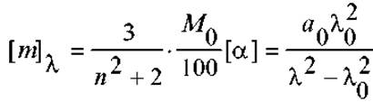
It agrees well with experimental data at wavelengths not too close to the absorption band of atoms or groups adjacent to the asymmetric carbon atom (λ0). Systems described by the single-term Drude equation exhibit simple optical dispersion curves. However, for many systems, including PROTEINS AND Peptides, the Drude equation is inadequate because so-called anomalous dispersion should be observed near λ0. In the Drude equation, this represents a discontinuity point (division by zero), as approaching it would cause optical activity to approach infinity. In reality, this merely reflects the Limitations of the Drude equation (similar, for instance, to Coulomb's or Newton's laws when r→0). In practice, optical activity increases, but its dependence on λ is quite specific (see Fig.), and the observed phenomenon is known as the Cotton effect.

Near the absorption band (λ=λ0), optical activity increases sharply, passes through a maximum, and drops precipitously. Then, changing sign, it passes through an equally deep minimum and flattens out (see Fig.). For proteins, the maximum optical activity is observed at 230 nm.
The Cotton effect is accounted for by the empirical two-term equation proposed by Moffitt and Yang:
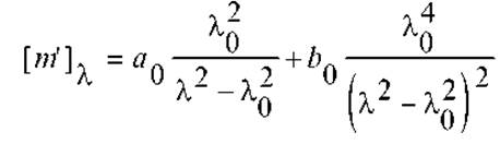
where [m’]λ is the reduced mean residue rotation.
The coefficients a0 and b0 are determined experimentally. To do this, the latter equation is rewritten as:
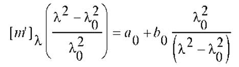
and plotted as a linear graph:
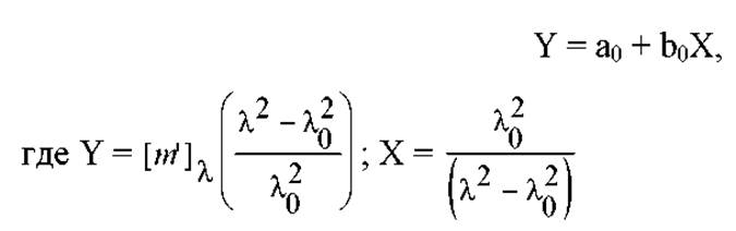
The figure shows, as an example, data plotted in linear coordinates for the optical rotatory dispersion of a synthetic peptide (λ0 = 212 nm). This peptide is a copolymer with the following composition: 5% L-Tyrosine and 95% L-glutamic acid (results obtained by Blout and Doty / or Blout et al. — *Note: 'Уорнес' is typically a typo for Blout in biochemistry literature, but keeping Translation natural*).
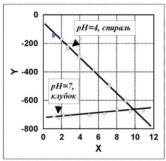
The parameter a0 reflects the rotation due to The Nature of The amino acid residues, while b0 reflects interactions within the α-helical segments of the peptide chain. The transition from a helical Structure to a random coil is accompanied by a decrease in the slope (b0). Right-handed helices in nonpolar Solvents are characterized by b0 values of approximately -630. For unordered structures in polar solvents, this value approaches zero (see Fig.).
Thus, observing the Cotton effect in proteins can serve as a diagnostic tool for their Secondary structure. It should be noted, however, that variations in b0 are significantly less pronounced in proteins than in synthetic peptides.
Last update: 06/08/2026
Editorial and Educational Adaptation: This material has been compiled based on the primary/original source text. The project team performed an editorial review, corrected technical inaccuracies, structured sections, and adapted the content for an educational format.
What was processed:
- elimination of formatting defects (OCR errors, structural breaks, corrupted characters);
- editorial organization of content;
- standardization of terminology in accordance with academic sources;
- verification of factual statements against the original source text.
All mentions of the author, publication year, and origin of the primary text have been preserved in accordance with the source.