Biochemistry - The Chemical Reactions of Living Cells, Volume 3 - D. Metzler 1980
Biochemical Genetics and the Synthesis of Nucleic Acids and Proteins
Genetic Methods
Mapping the Bacteriophage Chromosome
Much of our knowledge in biochemical genetics has been gained through The Study of Bacteriophages. Intensive research on the "T-even" phages T2, T4, and T6 began back in 1938 by Max Delbrück and his coworkers. Although the Viruses they studied were small, they turned out to be among the most complex known viruses (Box 4-D). The Genetic information contained in a single linear DNA molecule—which, in the case of phage T4, comprises 2∙105 Base Pairs—is sufficient to encode approximately 200 genes. The positions of 60 of these genes have been successfully mapped on the genetic map. Below, we briefly review the method used to achieve this.
Infection of an E. coli Cell by a bacteriophage occurs as follows: the phage injects its DNA through The Cell wall into the Cytoplasm. Approximately 20 minutes later, The Cell lyses, releasing about 100 fully formed copies of the original viral particle. This high rate of reproduction makes it possible to conduct genetic experiments in a test tube within 20 minutes that would require the entire population of the Earth if such experiments were performed on humans. The core principles underlying this method were clearly outlined by Benzer [130], who pioneered the fine-Structure mapping of a Gene. Bacteriophage particles, much like Bacteria, can be "plated" on an Agar petri dish. The only difference is that the agar must contain a uniform suspension of bacteria susceptible to the virus. Wherever the Viral Particles land on the plate, they infect a bacterium. The infection soon spreads to neighboring bacteria, resulting in The formation of a clear plaque (Fig. 15-20). The Number of viable viral particles in a suspension can be easily determined by counting the number of plaques formed upon plating.
Mutant bacteriophages can be detected in various ways, but the simplest approach is by the appearance of the plaques they form. Another easily identifiable Class of mutants consists of those with altered host range Specificity for certain bacterial strains. A key discovery that enabled the genetic mapping of bacteriophages was the finding that genetic recombination can occur between two phage particles within a host bacterium. Recombination can be illustrated as follows: two different mutant bacteriophage strains are grown in large quantities and mixed together in the presence of a large excess of bacteria. The progeny then reveal a certain number of viruses exhibiting the mutant traits of both strains, as well as an equal number of "wild-type" viruses. Although recombination between Mutations located close to each other on the DNA occurs rarely, its frequency still significantly exceeds the spontaneous mutation rate. Thus, while the experiment described above does not elucidate the precise Molecular Mechanism of recombination, it unequivocally demonstrates that recombination does occur.
The study of recombination frequencies between various phage strains soon revealed that certain mutation sites are tightly linked to one another, meaning recombination between them is rare. Other sites are loosely linked, and recombination between them occurs frequently. This situation is reminiscent of the observations made many years earlier with fruit flies (Drosophila)1), corn, and other higher organisms. The central premise underlying chromosome mapping in any Organism is the assumption that the recombination frequency between two mutations is directly proportional to the distance between their loci on the genetic map. For phage T4, a recombination frequency of 1% is adopted as the unit of map length. The entire T4 map spans 700 such units. The fact that this value exceeds 100% means that if genes are located at opposite ends of a chromosome, recombination between them can occur multiple times. In reality, a maximum crossover frequency of 50% is observed between genes that are far apart, and the linear relationship between recombination frequency and map distance holds true only for distances not exceeding 10 units [131].
How can a recombinant bacteriophage be detected rapidly? Benzer used two E. coli strains as host Cells: strain B and strain K. Bacteriophages carrying the mutant rII gene formed characteristic plaques on plates with strain B bacteria, but failed to grow on strain K bacteria. To determine the recombination frequency between two different rII mutants, the viruses were added to a liquid culture of B cells, within which they replicated. As a result of recombination occurring under these conditions, not only phages with both Selection/21.html">Types of mutations appeared, but also "standard" phages that had lost both mutations due to recombination. Because only recombinants of the latter type can grow on strain K cells, the presence of even a single recombinant can be identified among a billion progeny. Let us assume that the total length of the T4 phage DNA is 200,000 base pairs, meaning there are 286 base pairs per map unit. A recombination frequency of 0.01% implies that these mutations are separated by a mere three base pairs in the DNA chain. Based on this, Benzer concluded that even when mutations involve DNA bases located in the immediate vicinity of one another, the expected recombination frequency between them can be readily determined.
1) Chromosomal recombination in Drosophila via Crossing-over was first discovered in 1911 by Morgan and his coworkers.
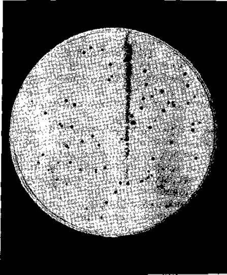
FIG. 15-20. Plaques formed by bacteriophage 11 growing on Staphylococcus aureus. Each clear, dark spot is formed as a result of bacterial lysis caused by the progeny of a single particle (photograph kindly provided by Pater Pattee).
For high-resolution genetic mapping to be practical, yet another method was required. A genetic map was constructed for a series of mutant bacteriophages carrying deletions spanning large segments of the rII gene. Using these mutants, it was possible to easily determine which specific region of the gene contained a given mutation. Subsequent recombination experiments using pre-identified mutations allowed for the precise localization of the mutation within the previously mapped gene segment. In this manner, Benzer successfully identified over 300 mutations in the rII gene. He concluded that the minimum distance between two mutant sites is in full agreement with the model of gene structure proposed by Watson and Crick.
Last update: 06/08/2026
Editorial and Educational Adaptation: This material has been compiled based on the primary/original source text. The project team performed an editorial review, corrected technical inaccuracies, structured sections, and adapted the content for an educational format.
What was processed:
- elimination of formatting defects (OCR errors, structural breaks, corrupted characters);
- editorial organization of content;
- standardization of terminology in accordance with academic sources;
- verification of factual statements against the original source text.
All mentions of the author, publication year, and origin of the primary text have been preserved in accordance with the source.