Biochemistry - The Chemical Reactions of Living Cells Volume 3 - D. Metzler 1980
Light in Biology
Vision
Light-Induced Transformations
Illumination of rhodopsin triggers a sequence of readily detectable spectral changes [133, 140]. The relaxation times indicated in the scheme (13-35) were obtained at 20°C.
Class="center">
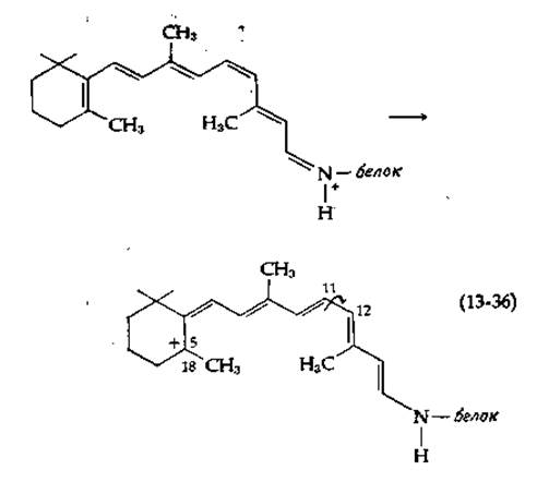
The initial transformation, recorded using laser flash photolysis and picosecond spectroscopy [141], takes place within 6∙10-12 s (6 ps). The absorption maximum of the resulting bathorhodopsin (prelumirhodopsin) is shifted toward longer wavelengths (bathochromic shift). This indicates an increased degree of conjugation, which is observed, for instance, during the isomerization of rhodopsin to all-trans-retinal [equation (13-34)]. However, it is unlikely that such a transformation could occur in so short a time, although the rapid formation of a strained all-trans-retinal is possible [141a]. A simple charge transfer yielding a carbonium ion1) [scheme (13-36)] cannot be ruled out either.
A subsequent, relatively slow rotation around the single bond indicated by the arrow can lead to The formation of the all-trans isomer, while the Cleavage of a proton from the C-18 atom results in a compound featuring an additional double bond between the C-5 and C-18 atoms. Exactly this Structure has been proposed for bathorhodopsin [141c]. Other possibilities under consideration include the formation of a biradical (in the triplet state) [142] as well as a charge-transfer complex involving a Tryptophan side chain [143].
1) Mathes and Stryer have recently provided direct evidence for charge transfer proceeding via mechanism (13-36) in the retinal Schiff base when the latter is in the first singlet excited state [141b].
The Nature of The primary photochemical stage, as well as the subsequent dark reactions, remains to be precisely elucidated [141, 144]. The sequence of reactions (13-35) can be halted at various stages by lowering the Temperature. Under specific conditions, an additional stage appears in this sequence. For instance, at 7 K, the first observable product is hypsorhodopsin, which absorbs at 437 nm (22 900 cm-1). The transition from metarhodopsin I to metarhodopsin II is of particular interest, as it represents the slowest stage that could still mediate the excitation of a Nerve Impulse (which travels along the rod to the synapse in approximately 1 ms). There is evidence that this step is accompanied by conformational changes. Current views on the Nature of the subsequent stages leading to the release of trans-retinal remain rather contradictory. Nevertheless, these steps are far too slow to play a significant role in the initiation of the nerve impulse.
What is the possible mechanism by which the sequence of reactions given in scheme (13-35) initiates a nerve impulse? The simplest assumption is that a conformational change within the retinal molecule during the isomerization of 11-cis-retinal to all-trans-retinal [scheme (13-34)] induces a protein conformation change, thereby conferring enzymatic activity upon the protein. Metarhodopsin II could potentially serve as the enzyme initiating the cascade of Chemical Reactions that culminates in a nerve impulse, though no experimental data currently support this hypothesis. Alternatively, the induced conformational changes within the protein molecule may open a channel in the disk membrane, allowing a certain substance to diffuse outward. Ca2+ is increasingly being considered as a plausible candidate for this substance. The distance from the disk membranes to Cell/30.html">The Plasma Membrane of the rod is short enough for the released substance to reach the plasma membrane (where the nerve impulse is actually excited) via diffusion.
In disk membranes, rhodopsin accounts for 80% (or even more) of the total protein, and it is possible that rhodopsin itself acts as the light-gated "pore" or "gate" [145]. The corresponding channel might pass directly through each rhodopsin molecule or run along the axis of the oligomeric aggregate. This hypothesis is supported by the observation that two-thirds of the peptide group hydrogen atoms in disk membranes apparently form Hydrogen Bonds with solvent Water molecules (as inferred from hydrogen-tritium exchange rates) [146]. This fact is particularly striking given the hydrophobic nature of rhodopsin and the fact that in typical Globular Proteins, only one-third of the peptide protons are capable of rapid exchange. All of the above can be explained by assuming that the majority of the rhodopsin peptide protons reside inside a water-filled channel, access to which is opened by the action of light on the Schiff base formed by the binding of retinal to the protein [146].
It is also conceivable that a cooperative signal-transmission process takes place between a rhodopsin molecule and another distally located protein that controls membrane permeability. One might even postulate that this cooperative process extends widely enough for a distinct physical signal to propagate along the disk membrane, reaching the disk edge and triggering a specific chemical signal near the plasma membrane.
It is well established that increasing the intensity of light falling on the retina leads to a rapid decrease in its sensitivity. This phenomenon may stem from the phosphorylation of a specific region of the rhodopsin molecule by opsin kinase, which acts specifically on bleached rhodopsin. Phosphorylated rhodopsin is apparently less permeable to Ca2+ than the "normal" form [147]. Furthermore, the bleaching of rhodopsin activates phosphodiesterase, which hydrolyzes cyclic GMP with high efficiency [148]. Thus, light exposure can trigger numerous secondary alterations associated with a drop in cGMP concentration.
Last update: 06/08/2026
Editorial and Educational Adaptation: This material has been compiled based on the primary/original source text. The project team performed an editorial review, corrected technical inaccuracies, structured sections, and adapted the content for an educational format.
What was processed:
- elimination of formatting defects (OCR errors, structural breaks, corrupted characters);
- editorial organization of content;
- standardization of terminology in accordance with academic sources;
- verification of factual statements against the original source text.
All mentions of the author, publication year, and origin of the primary text have been preserved in accordance with the source.