Biochemistry - Chemical Reactions in Living Cells, Volume 3 - D. Metzler 1980
Light in Biology
Vision
Chemical Nature of Chromophores in Visual Pigments
The chromophore of rhodopsin is 11-cis-retinal, which, upon exposure to light, is isomerized [scheme (13-34)] into all-trans-retinal. The latter dissociates from the protein moiety of the molecule—the opsin apoprotein—which can subsequently spontaneously recombine with 11-cis-retinal to regenerate rhodopsin.
Class="center">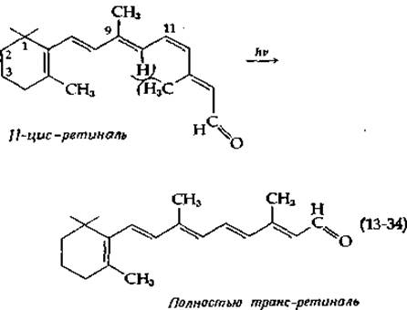
Other visual pigments related to rhodopsin are also known. Cone opsins complexed with 11-cis-retinal are called porphyropsins; they typically absorb light at slightly longer wavelengths than rhodopsin1). In certain freshwater species, the chromophore of the visual pigments (iodopsins) is 3-dehydroretinal. THE POSITION OF the absorption maximum of visual pigments depends on both The Nature of the bound aldehyde and the Nature of the protein, with the latter exerting a greater effect (Table 13-3). Thus, retinal-containing pigments absorb light over a wide wavelength range spanning from 467 to 528 nm (18,900–21,400 cm-1).
Table 13-3 Positions of the main absorption maxima of retinals and visual pigments
|
Free aldehyde |
Visual pigment |
||
|
Retinal (all-trans) |
387 nm 25,800 cm-1 |
||
|
Retinal (11-cis) |
376 nm 26,600 cm-1 |
Rhodopsin |
500 nm 20,000 cm-1 |
|
Porphyropsin (cones) |
522 nm 19,200 cm-1 |
||
|
3-Dehydroretinal (11-cis) |
393 nm 25,400 cm-1 |
Iodopsin |
562 nm 17,800 cm-1 |
When native rhodopsin is treated with sodium borohydride, the reduction reaction is relatively weak, but following protein bleaching, reduction proceeds rapidly and retinal becomes covalently bound to the protein to form a secondary amine. This indicates that in rhodopsin, retinal is linked to the protein moiety via a Schiff base. Attempts to identify the amino group to which this base is attached have yielded contradictory results.
1)However, in primates, Color Vision is apparently determined by three pigments with λmах = 447 nm (blue-violet region), 540 nm (green), and 577 nm (yellow); see MacNichol E. F., Jr, Sci. Am., 211, 48–56 (Dec. 1964). Note that one of the pigments absorbs light at shorter wavelengths than rhodopsin.
Some experiments pointed to the involvement of the ε-amino group of opsin, whereas others pointed to the amino group of phosphatidylethanolamine. Recently, by slowly reducing unbleached rhodopsin with cyanoborohydride, researchers successfully obtained a single product, the analysis of which led to the Conclusion that the Schiff base in the native pigment is formed at the Lysine amino group [133]. According to model system studies, the large bathochromic shift of the visual pigment absorption spectrum relative to that of free retinal is due to the presence of a strongly protonated Schiff base in the pigments and strong interactions between the polyene chain of retinal and the protein.
Last update: 06/08/2026
Editorial and Educational Adaptation: This material has been compiled based on the primary/original source text. The project team performed an editorial review, corrected technical inaccuracies, structured sections, and adapted the content for an educational format.
What was processed:
- elimination of formatting defects (OCR errors, structural breaks, corrupted characters);
- editorial organization of content;
- standardization of terminology in accordance with academic sources;
- verification of factual statements against the original source text.
All mentions of the author, publication year, and origin of the primary text have been preserved in accordance with the source.