Biochemistry: The Chemical Reactions of Living Cells, Volume 2 - D. Metzler 1980
How electrons meet oxygen, how ATP is generated in the process, and other related phenomena
Hemoproteins
Some names worth remembering
In 1879, the German physiological chemist Hoppe-Seyler discovered that two of nature's most vivid and striking colors share a common origin. He found that the red iron-containing heme in Blood and the green magnesium complex of chlorophyll a in leaves possess a similar ring Structure. However, it was Fischer, working in Munich, who provided rigorous proof for this relationship; between 1910 and 1940, he elucidated The structure of these compounds and established the nomenclature system and numbering scheme still in use today.
Porphins are large macrocyclic rings formed from four smaller pyrrole rings linked together by four methene bridges. In chlorins, which are found in chlorophylls, one of the rings (D) is in a reduced form. In a special Class of porphins, the Porphyrins, the periphery of the large ring bears eight substituent groups. Porphyrins, like chlorins and the corins of vitamin B12 (Supplement 8-L), are biosynthesized from porphobilinogen. This compound polymerizes via two pathways (Fig. 14-13) to yield type I and type III porphyrins (Fig. 10-1). In The formation of type I porphyrins, the polymerization of porphobilinogen proceeds in a regular manner, resulting in a symmetrical alternation of carboxymethyl and carboxyethyl side chains (often referred to as acetic and propionic acid side chains, respectively) around the ring. However, the biologically most important porphyrins belong to type III, in which the first three rings, A, B, and C, have the same sequence of carboxymethyl and carboxyethyl side chains as type I porphyrins, whereas ring D is inverted. As a result, the carboxyethyl side chains of rings C and D end up adjacent to each other (see Fig. 10-1). Porphyrins bearing four carboxymethyl and four carboxyethyl side groups are called uroporphyrins. Type I and III uroporphyrins are excreted in small amounts in the urine. Another excretion product is coproporphyrin III, in which all carboxymethyl side chains are decarboxylated to methyl groups. The coloration of the feathers of the tropical turaco bird is due to a copper(II)-containing complex of coproporphyrin III; this porphyrin, along with others, is also found in bird eggs.
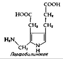
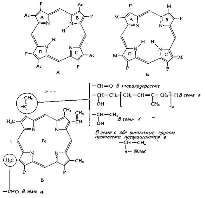
FIG. 10-1. Structures of several biologically important porphyrins. A. Uroporphyrin I; Ac =—CH2COOH and P =—CH2CH2COOH. B. Coproporphyrin III (Note that, compared to A, a different tautomeric form of the ring is depicted here; such Tautomerism is characteristic of all porphyrins). C. Protoheme — the Fe2+ complex of protoporphyrin IX, present in Hemoglobin, Cytochromes b, and various other Proteins.
All Hemoproteins are derived from protoporphyrin IX, which is the decarboxylation product of coproporphyrin III; in place of two carboxyethyl side chains, it contains two vinyl groups (Fig. 10-1).
Last update: 06/08/2026
Editorial and Educational Adaptation: This material has been compiled based on the primary/original source text. The project team performed an editorial review, corrected technical inaccuracies, structured sections, and adapted the content for an educational format.
What was processed:
- elimination of formatting defects (OCR errors, structural breaks, corrupted characters);
- editorial organization of content;
- standardization of terminology in accordance with academic sources;
- verification of factual statements against the original source text.
All mentions of the author, publication year, and origin of the primary text have been preserved in accordance with the source.