BIOCHEMISTRY - Main Regulators and Biological Fluids of the Human Body - 2016
3. NUCLEIC ACIDS
3.2. Chemical Composition of Nucleotides
Nucleic Acids are heteropolymers whose monomers are NUCLEOTIDES.
Nucleotides are Organic compounds consisting of a nitrogenous base, a carbohydrate residue, and a phosphoric acid residue.
3.2.1 Nitrogenous Bases
Nitrogenous bases are aromatic heterocyclic compounds derived from purine or pyrimidine.
Class="center">
Purine bases (Purines) are adenine and guanine.
Pyrimidine bases (Pyrimidines) include cytosine, thymine, and uracil.
The purine bases adenine (Ade) and guanine (Gua), as well as the pyrimidine base cytosine (Cyt), are constituents of both DNA and RNA. The pyrimidine base thymine (Thy) is exclusively found in DNA. The pyrimidine base uracil (Ura) is exclusively found in RNA.
3.2.2. Carbohydrate Components
The carbohydrate moiety of nucleic acids is represented by Monosaccharides (pentoses) — deoxyribose in DNA and ribose in RNA. They always exist in the β - D -furanose form.

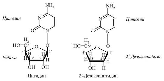
Unlike ribose, deoxyribose has a hydrogen atom (-H) instead of a hydroxyl group (-OH) at the second carbon atom, which increases the Stability of the DNA molecule.
3.2.3. Nucleosides
Compounds of nitrogenous bases with carbohydrate residues form nucleosides, which are named According to the specific nitrogenous bases and pentoses they contain (Table 3)
Composition and Names of Nucleosides
Nucleoside |
Designation |
Nitrogenous Base |
Pentose |
Adenosine |
A |
Adenine |
Ribose |
Deoxyadenosine |
dA |
Adenine |
Deoxyribose |
Guanosine |
G |
Guanine |
Ribose |
Deoxyguanosine |
dG |
Guanine |
Deoxyribose |
Cytidine |
C |
Cytosine |
Ribose |
Deoxycytidine |
dC |
Cytosine |
Deoxyribose |
Uridine |
U |
Uracil |
Ribose |
Thymidine |
T |
Thymine |
Deoxyribose |
Thus, for example, adenine and ribose form the nucleoside adenosine (abbreviated as A). Corresponding derivatives of other nitrogenous bases are named guanosine (G), uridine (U), thymidine (T), cytidine (C). If the carbohydrate residue is represented by 2-deoxyribose, a deoxynucleoside is formed, for example, 2'-deoxyadenosine (dA).
Below are the structural formulas of RNA nucleosides — adenosine and cytidine, and DNA nucleosides — 2'-deoxyadenosine and 2'-deoxycytidine.

3.2.4. Mononucleotides
In Cells, the 5'- OH group of the nucleoside carbohydrate residue is esterified with phosphoric acid. Monophosphate esters of nucleosides are called mononucleotides. For example, the structural formulas of the nucleotides guanosine monophosphate (GMP) and 2'-deoxythymidine monophosphate (dTMP) are shown below.
Mononucleotides are the structural units of nucleic acids.

If the 5'-phosphate group of a mononucleotide binds with another phosphoric acid residue, a nucleoside diphosphate is formed, and with two residues—a nucleoside triphosphate. Below are the structural formulas of adenosine diphosphate (ADP or ABR) and adenosine triphosphate (ATP or ATR).

ADP (ABR) and ATP (ATR) are essential Coenzymes in METABOLISM/26.html">Energy Metabolism.
3.2.5. Structure of Nucleic Acids
The nucleic acid molecules of all types of Living organisms are long, unbranched polymers of mononucleotides. In nucleic acid molecules, nucleotides are linked via the phosphoric acid residue of one nucleotide and the 3'-OH group of the sugar of another. Thus, mononucleotides are joined within the nucleic acid molecule by a phosphodiester bond.
Polynucleotides composed of ribonucleotide units are called Ribonucleic Acids—RNA, while those composed of deoxyribonucleotide monomers are called Deoxyribonucleic Acids—DNA (Fig. 11).
Fig. 11. Structure of RNA and DNA
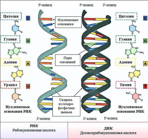
When designating polynucleotides, the abbreviated names of the nucleoside units are indicated in the direction from the 5'-end of the nucleic acid to the 3'-end (5' —> 3').
Thus, the beginning of the RNA in Fig. 11 can be written as UCCUA.C, etc.
3.2.6. Deoxyribonucleic Acids (DNA)
The Introduction/19.html">Primary Structure of DNA is a heteropolymer double strand of nucleotides. The DNA molecule is twisted into a double helix, the strands of which are connected to each other along their entire length by Hydrogen Bonds. Hydrogen bonds form between the nitrogenous bases located in the interior of the molecule. Nitrogenous bases pair according to THE PRINCIPLE OF complementarity: A – T, G – C. The A–T pair can form two, and the G–C pair three linear and therefore stable hydrogen bridges (hydrogen bonds). Base pairing in DNA is shown below.

Functionally, the two DNA strands are not equivalent. The coding strand (template, sense strand) is the one that is read during Transcription. This very strand serves as a template for RNA. The non-coding strand (antisense) is similar in sequence to RNA (provided T is replaced by U).
The model of the molecular structure (Spatial Structure) of DNA was proposed by J. Watson and F. Crick in 1953, for which they were awarded the Nobel Prize. The model has been fully confirmed experimentally, and this discovery played an exceptionally important role in The Development of molecular biology and genetics.
Despite differences in DNA Structure, the total Nucleotide Composition of all types of DNA shares common regularities established by E. Chargaff (Chargaff's rules):
1) the molar ratio of A to T is equal to 1 (A/T = 1);
2) the molar ratio of G to C is equal to 1 (G/C = 1);
3) the sum of purine bases is equal to the sum of pyrimidine bases.
3.2.7. Ribonucleic Acids (RNA)
The primary structure of RNA is a heteropolymer strand consisting of nucleotides (A, G, C, U) whose sugar component is ribose.
Depending on their function, RNAs are subdivided into three types:
- Messenger RNA – mRNA (or template RNA – mRNA),
- ribosomal - rRNA
- transfer - tRNA.
Each type of RNA has its own size, structure, and lifespan (Table 4).
Table 4
Classification of RNA
Characteristic |
Type |
||
mRNA |
rRNA |
tRNA |
|
Percentage of total RNA, % |
5 |
80 |
10 - 20 |
Size, nt |
400 - 6000 |
120-5000 |
70 - 90 |
Structure |
Single strand |
Associated with Proteins |
Cloverleaf shape |
Number of subtypes |
1000 |
4 |
Over 50 |
Lifespan |
Short |
Long |
Long |
Nucleus, Cytoplasm, Ribosomes, Cell/35.html">Mitochondria, Plastids |
Ribosomes |
Cytoplasm, ribosomes |
|
Function |
Carries Genetic information from the Cell Nucleus to the cytoplasm to the site of Protein Synthesis |
Structural component of ribosomes - forms the ribosome |
Transports Amino Acids to the site of protein synthesis (to the ribosome) |
All Three types of RNA are involved in Protein Biosynthesis - Translation (see subsection 3.6 below).
Last update: 06/08/2026
Editorial and Educational Adaptation: This material has been compiled based on the primary/original source text. The project team performed an editorial review, corrected technical inaccuracies, structured sections, and adapted the content for an educational format.
What was processed:
- elimination of formatting defects (OCR errors, structural breaks, corrupted characters);
- editorial organization of content;
- standardization of terminology in accordance with academic sources;
- verification of factual statements against the original source text.
All mentions of the author, publication year, and origin of the primary text have been preserved in accordance with the source.