Amino Acids, Peptides and Proteins - Dévényi T., Gergely J. 1976
Protein Chromatography
Protein chromatography on a DEAE-cellulose column
Serum protein chromatography on a DEAE-cellulose column
a) Preparation of DEAE-Cellulose. The required amount of DEAE-cellulose is first washed three times with distilled Water by decantation, and then sequentially with the following solutions: 0.5 M NaOH, distilled water, 0.3 M NaH2PО4, distilled water, 0.5 M NaOH, distilled water, and 0.005 M Na2HPО4.
Washing of the ion exchanger with distilled water is always continued until the pH of the decanted water equals the pH of the added water. In the final washing cycle, the DEAE-cellulose is equilibrated with the starting buffer solution.
Class="center">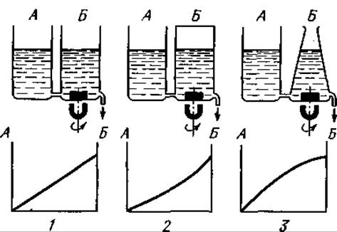
Fig. 41. Gradient elution in Ion-exchange Chromatography. The buffer solution filling reservoir A has a higher Ionic strength than the solution in mixer B. Concentration gradients: 1 — linear; 2 — “concave”; 3 — “convex”.
b) Column packing. Packing the column and compacting the ion exchanger are performed using compressed air. The ion exchanger is suspended in the starting buffer solution, stirred to obtain a homogeneous suspension, poured into a 1x60 cm column, and allowed to settle. The cellulose is then compacted with compressed air at a pressure of 0.5 atm. After this, the completeness of equilibration is checked, i.e., whether the pH of the eluate equals the pH of the starting buffer solution. If they are not equal, additional starting buffer solution is passed through the ion exchanger column until the column is equilibrated.
c) Preparation for gradient elution. A schematic diagram of the vessels used, as well as the types of gradients, is shown in Fig. 41. The volume of the mixer, which can be any round vessel, is 500 ml, and the volume of the buffer reservoir is 1000 ml. The reservoir is placed 7.5 cm above the mixer. The mixer is filled with 0.01 M phosphate buffer solution, pH 8.4, and the reservoir is filled with 0.3 M buffer solution, pH 4.2. The starting 0.005 M phosphate buffer solution, pH 8.4, is applied to the column from a separate vessel.
d) Sample application. 2 ml of fresh or thawed serum is dialyzed against the starting buffer solution for 24 hours. The buffer solution above the ion exchanger in the column is carefully removed, and the dialyzed serum is applied with a pipette. After application, any remaining sample is washed down three times with separate 2 ml portions of the buffer solution.
e) Chromatography. Chromatography is initially carried out in the starting buffer solution at a flow rate of 50 ml/h until the first peak emerges. Then, without changing this flow rate, gradient elution is started. Eluate fractions of 3–5 ml are collected (about 500 ml in total), and the protein content in the collected fractions is determined either by continuous spectrophotometry, for example, using an LKB “Uvicord”, or by measuring the absorbance of each fraction at 280 nm.
NOTES
1. Serum chromatography can be performed at room Temperature, but it is still preferable to cool the column.
2. According to our data, the capacity of different DEAE-cellulose preparations can vary significantly and may also change after regeneration. Therefore, in each case, columns of different sizes must be used and different amounts of protein applied. Consequently, it is recommended to determine the most suitable chromatographic conditions in a preliminary experiment with the specific batch of ion exchanger.
3. DEAE-cellulose is regenerated in the same way as during its preparation for serum fractionation (see p. 209).
4. The easiest way to identify the serum protein components in different eluate fractions is by Immunoelectrophoresis. The first peak emerging with the starting buffer solution usually contains a significant portion of serum IgG; this is followed by the remaining γ-globulin fractions, β- and α-globulins, with albumin being eluted last.
5. If gradient elution is performed as described by Fahey, 0.1 M phosphate buffer solution, pH 8.0, is poured into the mixer, and 0.3 M phosphate buffer solution, pH 8.0, is poured into the reservoir.
6. Continuous delivery of the buffer solution at a constant rate is best achieved using suitable pumps. LKB pumps used for this purpose, such as “Miniflow”, “Peristaltic”, and “Perpex”, have proven to be highly reliable.
7. Concentration of protein fractions by pressure dialysis. For this purpose, dialysis membrane tubing with a diameter of 8 mm is used (“Visking” tubing from Scientific Instrument Centre Ltd). A piece of tubing of appropriate length is moistened with distilled water, and one end is tied with a double knot. To check the tightness of the bottom knot, the tubing is immersed in water and inflated with air. The upper end of the dialysis tubing is passed through a hole in a rubber stopper and fitted onto the stem of a funnel of appropriate size; the funnel with the attached dialysis tubing is then inserted into the hole of the rubber stopper. The protein solution to be concentrated is poured through the funnel into the dialysis tubing, air bubbles are expelled by gentle squeezing, and the filled tubing is placed in a Buchner filter flask of suitable size so that the stopper, through which the funnel stem passes, fits tightly into the neck of the flask. The tightness of the rubber-to-Glass seal can be checked by applying a few drops of water around the edge of the stopper.
The filter flask is connected to a water aspirator to create a vacuum. Under vacuum, the solvent filters through the dialysis membrane into the flask, and concentration of the protein solution begins. A clamp is placed on the outlet hose of the filter flask to maintain the vacuum, and the entire system is placed in a refrigerator or a cold room. If the volume of the solution to be concentrated is larger than the capacity of the funnel, more protein solution is added as concentration proceeds. When the volume of the solution has decreased to the desired level, the filter flask with the dialysis tubing is removed from the refrigerator, the clamp is opened, and the tubing is taken out of the flask. The membrane is then cut open, and the concentrated protein solution is transferred to an appropriate vessel, rinsing the remaining solution from the walls of the dialysis tubing with a small amount of buffer.
8. Concentration of solutions using Lyphogel. Lyphogel, manufactured by Gelman Instrument Company (USA), is a polyacrylamide gel used to concentrate solutions of high-molecular-weight substances. Each gram of Lyphogel can bind up to 5 g of water, with equilibrium being established within 5 h. The solution to be concentrated is placed in a vessel of appropriate size, and enough Lyphogel is added to achieve the required concentration (keeping in mind that 1 g of Lyphogel, when swollen in an aqueous medium, occupies a volume of 25 ml). When concentration is complete, the swollen Lyphogel is easily removed by filtration. After drying at 65°C, it can be reused1.
1 One of the fastest and most convenient Methods for concentrating protein solutions is Membrane filtration (see p. 27). — Ed. note.
Last update: 06/08/2026
Editorial and Educational Adaptation: This material has been compiled based on the primary/original source text. The project team performed an editorial review, corrected technical inaccuracies, structured sections, and adapted the content for an educational format.
What was processed:
- elimination of formatting defects (OCR errors, structural breaks, corrupted characters);
- editorial organization of content;
- standardization of terminology in accordance with academic sources;
- verification of factual statements against the original source text.
All mentions of the author, publication year, and origin of the primary text have been preserved in accordance with the source.