INVERTEBRATE ZOOLOGY IN THREE VOLUMES - BOOK 2 - H.Y. Shcherbak - 1996
PHYLUM ARTHROPODA
SUBPHYLUM TRILOBITOMORPHA
CLASS TRILOBITA
The DORSAL SIDE OF the trilobite body is covered by a hard exoskeleton—a dorsal shield impregnated with calcium carbonate and sometimes calcium phosphate. The surface of the carapace often features various sculptural patterns.
The dorsal shield is typically elongate-oval in shape and divided by two longitudinal furrows into three parts, or lobes; the middle lobe is more convex. In addition to this longitudinal division, the carapace is also transversely divided into cephalic (HEAD), thoracic, and pygidial (tail) shields (Fig. 182). The tripartite division gives the Class its Latin name (tri — three, lobus — lobe).
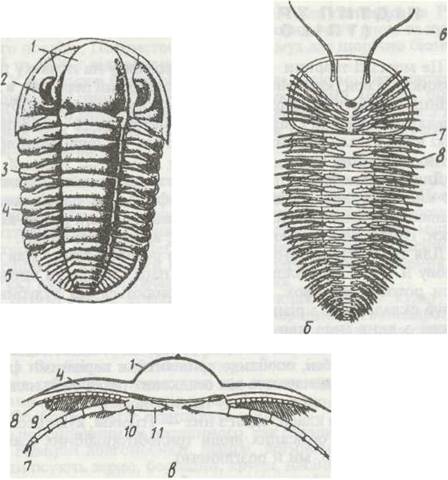
Fig. 182. Diagram of trilobite Structure:
a, b — general appearance from the dorsal and ventral sides, respectively; c — cross-section of the trunk; 1 — median region of the cephalic shield; 2 — compound eye; 3, 4 — median and lateral Regions of the thoracic shield; 5 — pygidial shield; 6 — antenna; 7 — appendage; 8 — respiratory appendage (epipodite); 9 — branchial lamellae; 10 — basal segment of the appendage; 11 — gnathobase
On the sides of the cephalic shield, most trilobites possess variably developed eyes of two types: faceted and aggregated. Faceted eyes consist of A large number of relatively large hexagonal or quadrangular lenses (facets, or ommatidia) covered by a common membrane, with the number of facets ranging from several dozen to 15,000. Aggregated eyes consist of rounded lenses separated from one another by interstices of cuticular meshwork. The number of facets in these eyes ranges from one or two to several hundred. In addition to these eyes, paired simple ocelli ("median eye") have been found in the middle of the cephalic shield in many species; another pair of similar eyes was sometimes located on the anterior margin of the head shield. Completely blind forms are also known.
In contrast to the dorsal side, the ventral side is flat and covered by a thin membrane. On the underside of the head, anterior to the Mouth opening, is a pair of multi-segmented antennae; four pairs of mouth appendages are structurally identical to the trunk appendages. All of them are uniramous, with a dorsal branchial outgrowth (epipodite); their basal segments bear a specialized gnathobase directed toward the midline of the body. Consequently, in addition to locomotion, all trilobite legs also served respiratory and masticatory Functions.
The internal anatomy of trilobites is poorly studied. In some fossil trilobite remains, impressions of certain Organs can be found on the inner side of the dorsal shield. The mouth was located on the underside of the head, followed by a short Esophagus that opened into a Stomach, which was also housed in the cephalic region. Short blind caeca extended in all directions from The Stomach; these structures are believed to be homologous to the digestive gland of crustaceans. Further back, the tube-like intestine extended to the pygidium, where it terminated in the anus. A long canal running above the intestine is considered to be a remnant of the circulatory
system.
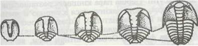
Fig. 183. Successive stages of postembryonic development in trilobites, starting with the protaspis
Trilobites are gonochoric (dioecious). A certain degree of Sexual Dimorphism was observed in The structure of males and females. Development was accompanied by metamorphosis comprising several phases. Trilobites laid small eggs (their structure and Cleavage type remain unknown), which hatched into a larval stage known as the protaspis (Fig. 183). Initially, the larva possessed an undivided body with two longitudinal furrows; subsequently, it simultaneously divided into a cephalic lobe and an anal lobe with several segments in between. The size of the protaspis did not exceed 1 mm. Thereafter, with each molt, the number of trunk segments increased; the growth zone for new segments was located just anterior to the pygidium. Fully segmented individuals continued to grow for some time without changing their segment count. As a result, the adult, sexually mature individual was formed.
Most trilobites lived at shallow depths and led a benthic lifestyle, as evidenced by their flattened bodies, upward-directed eyes, lack of swimming appendages, and other features. Some were pelagic (free-swimming) animals: their eyes were directed laterally, and various bodily outgrowths helped maintain buoyancy in the Water Column. Deep-water trilobites were characterized by large eyes, whereas burrowing forms were blind. Larvae led a planktonic lifestyle and served a dispersal function.
The evolutionary zenith of trilobites occurred In the second half of the Cambrian and the Ordovician periods, after which their species diversity sharply declined; the last trilobites became extinct in the middle of the Permian period.
Last update: 13/08/2026
Editorial and Educational Adaptation: This material has been compiled based on the primary/original source text. The project team performed an editorial review, corrected technical inaccuracies, structured sections, and adapted the content for an educational format.
What was processed:
- elimination of formatting defects (OCR errors, structural breaks, corrupted characters);
- editorial organization of content;
- standardization of terminology in accordance with academic sources;
- verification of factual statements against the original source text.
All mentions of the author, publication year, and origin of the primary text have been preserved in accordance with the source.