IMMUNOLOGY - Roitt I. - 2000
Chapter 20. Tumor Immunology
HUMAN IMMUNE RESPONSES TO TUMORS AND MECHANISMS OF TUMOR EVASION
Most tumors are infiltrated by lymphoid Cells
Histological analysis of human tumors frequently reveals that they are infiltrated by inflammatory cells (Fig. 20.8). Lymphocytes and macrophages typically dominate the infiltrate, though other Cell types may also be present, including dendritic cells, granulocytes, and mast cells. A more precise analysis of tumor-infiltrating cell types using Monoclonal Antibodies (mAbs) to identify lymphoid cell subpopulations (Fig. 20.9) demonstrates that most major lymphocyte subsets can be present within tumors.
Class="center">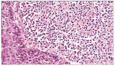
Fig. 20.8. Immunological reaction in breast Cancer. Section of a tumor surrounded by a dense infiltrate of mononuclear cells. The inflammatory response indicates that tumor cells are recognized by The Immune System, which has the potential to slow tumor growth or destroy tumor cells. Hematoxylin and eosin staining.

Fig. 20.9. CD4+ and CD8+ T cells in a breast cancer tumor. These cells were identified using an alkaline phosphatase immunostaining method (pink) with monoclonal antibodies. Sections are counterstained with hematoxylin. Surrounding CD4+ cells (top) and fewer CD8+ cells (bottom) are visible, along with occasional intra-tumoral lymphocytes.
Monoclonal antibodies specific for the IL-2 receptor, MHC class II Antigens, and other activation markers can also be used to determine the level of cellular activation. However, no clear-cut associations have yet been established between specific lymphoid subpopulations and malignancy prognosis. This may be because only a small fraction of the infiltrating cells are actually tumor-specific. Several studies have been undertaken to investigate the in vitro Functions of tumor-infiltrating lymphocytes; these are discussed in the next section.
Antitumor responses detected in mixed lymphocyte-tumor cell cultures
The concept that antigen-primed Th and Tc cell responses can be detected by restimulation with specific antigen in vitro was tested in the following experiments. To determine whether a patient's immune system can mount a response against their tumor, patient lymphocytes were stimulated with inactivated tumor cells in mixed lymphocyte-tumor cell cultures (MLTC) (Fig. 20.10). Lymphocytes for this purpose can be sourced from peripheral Blood, tumor-draining Lymph Nodes, or the tumor itself (the latter termed tumor-infiltrating lymphocytes, or TILs).

Fig. 20.10. Lymphocytes isolated from blood, tumor-draining lymph nodes, or directly from tumor tissue were co-cultured with autologous tumor cells inactivated by X-irradiation or mitomycin C Treatment. The proliferative capacity of the lymphocytes was then determined (by 3H-thymidine incorporation). An isotope-release assay was also developed to measure the ability of lymphocytes to lyse target cells.
Such mixed cultures have been shown to stimulate CD4+ Th cells, resulting in their proliferation and the release of effector cytokines. CD8+ Tc cells are also generated in these cultures, and their cytotoxic activity can be measured using a 51Cr-release assay (see Fig. 29.26). Tc cells can also be obtained by culturing lymphocytes in the presence of IL-2 to expand the effector cell population generated in vivo. Tc cell activity must be distinguished from natural killer (NK) cell activity; to differentiate them, the expanded lymphocyte population is tested using appropriate standard cultures.
Multiple T-cell specificities are generated in MLTC
Th and Tc cell specificities are similar to one another and exhibit varying reactivity profiles against different target cells in individual patients (Fig. 20.11).
✵ In both cases, only a minor fraction of the cloned T cells is specific for the autologous tumor.
✵ Some clones react with the autologous tumor as well as with all or part of the tested autologous Tissues.
✵ A number of clones show reactivity against autologous and certain allogeneic tumors.
✵ The remaining cells exhibit Specificity for many tumor target cells.

Fig. 20.11. Lymphocytes harvested from MLTC were evaluated in a 51Cr-isotope release assay using various tumor and normal cell types (isotope release into the culture medium indicates cell death). Lymphocytes from different patients differ in specificity; their properties range from The ability to lyse exclusively autologous tumor cells to the nonspecific lysis of all target cells. In the latter case, activated NK cells or T cells may be mediating the effect. Among T lymphocytes, CD4+ and CD8+ populations function similarly, displaying a comparable diversity of responses.
Tumor-specific T cells are found in patients with various types of malignancies. Melanoma appears to be particularly immunogenic. Several melanoma-associated antigens (MAAs) recognized by murine monoclonal antibodies have been identified. MAAs expressed by certain normal cells, along with tyrosinase (a pigment cell-associated enzyme), serve as targets for Tc cells. No Mutations have been detected in either MAA genes or the tyrosinase Gene in tumor cells. Tumor-bearing patients also mount a response against the mutant ras oncogene product and the tumor suppressor gene product p53.
A third type of T-cell clone has been isolated from breast and Ovarian cancer patients. Recent evidence indicates that these lines are capable of responding specifically, yet in a non-restricted manner, to a repeating core peptide of mucin. It remains unclear whether this peptide is presented in complex with MHC molecules or via an alternative mechanism (e.g., acting as a superantigen). Mucin-responsive cells belong to either Th or Tc lymphocyte subsets (CD4+ or CD8+).
The in vivo significance of cytotoxic responses detected in vitro remains unclear, although in animal model experiments, cultured antitumor cytolytic cells can induce tumor regression.
Tumors are able to evade the Immune Response through multiple pathways
Spontaneously developing tumors are capable of progressive growth leading to the death of the Organism; consequently, in many cases they successfully evade the immune response. Various hypotheses have been proposed to explain this phenomenon. The most obvious assumption is that tumors are non-immunogenic. The cause of non-immunogenicity may not be the absence of tumor-specific antigens, but rather their inadequate presentation on tumor cells. The induction of an immune response requires costimulatory factors, which can be cell surface molecules or cytokines secreted by antigen-presenting cells (APCs). The B7 molecule, found on specialized APCs, is now known to serve as a key costimulatory factor; it acts by binding to the CD28 marker On the surface of T cells (see Chapter 11). In experiments, the presentation of MHC molecule–antigenic peptide complexes to T-cell receptors in the absence of B7 can induce anergy. It has long been known that immune responses are suppressed in cancer patients. Recent studies of peripheral blood lymphocytes from such patients have revealed impaired T-cell signal Transduction; tumor-infiltrating lymphocytes may, in some cases, be anergic. Furthermore, if tumor cells lack class II MHC antigens, the initiation of a Th-cell response depends on the Processing of tumor antigens by specialized APCs (Fig. 20.12).

Fig. 20.12. Tumor antigens can be presented to T cells in various ways. 1. Direct antigen presentation in the absence of costimulatory molecules, leading to anergy. 2. Direct antigen presentation by tumor cells expressing costimulatory molecules, leading to Tc-cell activation. 3. Direct presentation on tumor cells and indirect presentation by specialized APCs, leading to the activation of both Tc and Th cells.
Tumor cells may also lack other molecules, such as LFA-1, LFA-3, or ICAM-1, which are required for lymphocyte adhesion (see Chapter 5), or they may express anti-adhesive molecules such as mucin. Tumor cells are also capable of secreting immunosuppressive cytokines, such as TGF-β. A particularly important mechanism of tumor immune evasion is the loss of MHC antigens, resulting in an inability to present tumor antigen Peptides. In more than 50% of cases, tumors lose one or more class II MHC alleles, and sometimes all of these alleles (Fig. 20.13).

Fig. 20.13. Breast cancer tissue reacting with monoclonal antibodies against a monomorphic determinant of class I HLA antigens. Only stromal cells are stained (brown), because malignant epithelial cells lose the ability to express normal class I MHC antigens. Approximately 50% of primary human malignant tumors are of epithelial origin. Aberrant expression of class II MHC antigens occurs on the cells of certain tumors. (Indirect immunoperoxidase staining method; hematoxylin counterstaining.)
Last update: 13/08/2026
Editorial and Educational Adaptation: This material has been compiled based on the primary/original source text. The project team performed an editorial review, corrected technical inaccuracies, structured sections, and adapted the content for an educational format.
What was processed:
- elimination of formatting defects (OCR errors, structural breaks, corrupted characters);
- editorial organization of content;
- standardization of terminology in accordance with academic sources;
- verification of factual statements against the original source text.
All mentions of the author, publication year, and origin of the primary text have been preserved in accordance with the source.