IMMUNOLOGY - Roit A. - Mir 2000
Chapter 20. Tumor Immunology
TUMOR-ASSOCIATED ANTIGENS RECOGNIZED BY ANTIBODIES
Some Antigens are unique to tumors
Numerous studies aimed at identifying tumor-specific antigens have employed either serum from the tumor-bearing animal itself (autologous typing) or sera from animals immunized with tumor tissue (heterologous typing). More recently, investigations have begun using Monoclonal Antibodies derived from autologous or heterologous B Cells. While definitive evidence for true tumor-specific antigens remains sparse, several types of antigens associated with tumors in various ways have been successfully identified.
Sera from Cancer patients reveal widely distributed antigens
When using sera from cancer patients or monoclonal antibodies raised against their tumor cells, researchers typically detect antigens that are broadly expressed On the surface—or, more frequently, in the interior—of both tumor and normal cells. The corresponding antibodies largely belong to the IgM Class and consequently exhibit low affinity. Similar monoclonal antibodies can be generated using immortalized B cells from healthy individuals. These antibodies recognize autoantigens, and their role, if any, in the body's Immune Response to tumors remains unclear.
Tumor Immunology
Tumors may express normal differentiation antigens characteristic of rare normal Cell types
Most tumor cells represent a clone—the progeny of a single cell—and cells of that specific Lineage may be relatively scarce in the body. Consequently, tumor cells express antigens that are normally present only in select types of normal cells. Examples of such antigens include the common acute lymphoblastic leukemia antigen (CALLA, or CD10) (Fig. 20.7) and carcinoembryonic antigens (CEA), which function as differentiation antigens expressed during embryonic development but typically absent (or expressed at extremely low levels) in adult Tissues.
CEAs include α-fetoprotein (AFP), produced by Liver tumor cells, and antigens produced by human colon carcinoma cells and other epithelial tumors.
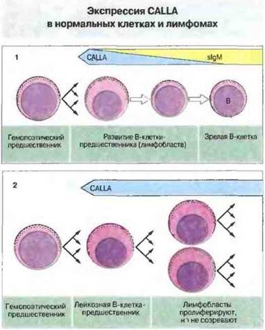
Fig. 20.7.
1. CALLA is normally expressed only on precursor B cells (lymphoblasts), which account for less than 1% of normal Bone Marrow cells. 2. CALLA expression is significantly upregulated in the most common form of childhood leukemia.
Normal cellular antigens expressed by tumors can be altered through glycosylation
Alterations in the glycosylation of cellular molecules are a hallmark of many tumors. These changes can lead to the expression of novel carbohydrate epitopes, such as the Thomsen-Friedenreich antigen—a disaccharide not typically detected on normal cells. Aberrant Blood Group Antigens can arise in a similar fashion. Furthermore, altered glycosylation can expose protein backbone epitopes that are rarely accessible in normal cells. A prime example is provided by polymorphic epithelial mucins produced by many normal epithelial cells. These are high-molecular-weight Glycoproteins in which a repeating core peptide carries carbohydrate side chains. In epithelial tumors, a novel protein epitope is revealed within this core Structure.
Last update: 13/08/2026
Editorial and Educational Adaptation: This material has been compiled based on the primary/original source text. The project team performed an editorial review, corrected technical inaccuracies, structured sections, and adapted the content for an educational format.
What was processed:
- elimination of formatting defects (OCR errors, structural breaks, corrupted characters);
- editorial organization of content;
- standardization of terminology in accordance with academic sources;
- verification of factual statements against the original source text.
All mentions of the author, publication year, and origin of the primary text have been preserved in accordance with the source.