Review of Medical Physiology - William F. Ganong 2002
Endocrine System, Metabolism, and Reproduction
Sex Differentiation and Development
Sex Differentiation and Development - Aberrant Sexual Differentiation
Given the above, it can be expected that DISORDERS OF SEXUAL development are caused by genetic or hormonal abnormalities, as well as other non-specific teratogenic factors. The main groups of such disorders are summarized in Table 23-1. A specific defect in gametogenesis is chromosomal nondisjunction—a phenomenon in which a pair of Chromosomes fails to separate, causing both chromosomes to end up in one of the daughter Cells during Meiosis. The four abnormal zygotes that can form due to nondisjunction of one of the X chromosomes during oogenesis are shown in Fig. 23-8. In individuals with an XO chromosomal Complement, the Gonads are rudimentary or absent, while the External female genitalia are developed. They are of short stature, often present with other Congenital Malformations, and fail to undergo Puberty at the appropriate time. This syndrome is referred to as gonadal dysgenesis, ovarian agenesis, or Turner syndrome.
Class="center">Table 23-1. Classification of major disorders of sexual differentiation in humans. Many of these syndromes can present with varying forms depending on the degree and manifestations.

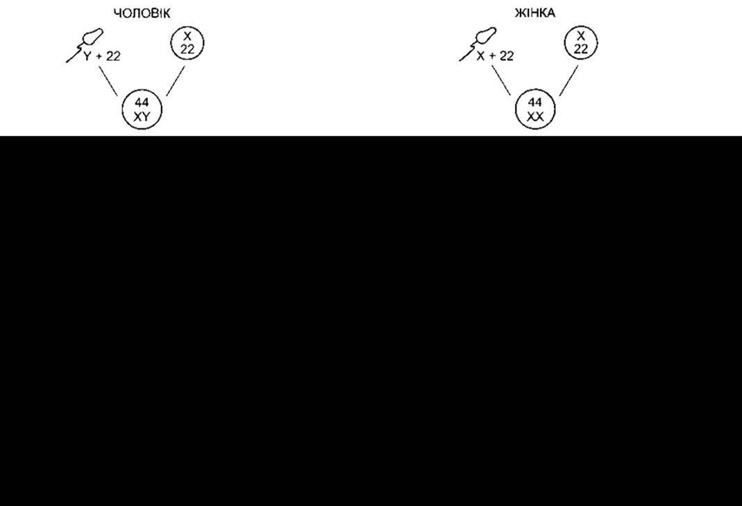
Fig. 23-5. Normal human Sex Determination, differentiation, and development (schematic representation); MIF – Müllerian-inhibiting factor; T – testosterone; DHT – dihydrotestosterone.
Individuals with an XXY complement—the most common sex chromosome disorder—have normal male external genitalia. During puberty, their testosterone secretion is entirely sufficient for The Development of male characteristics, yet abnormal seminiferous tubules are formed, and intellectual disability is more frequent among them. This syndrome is known as seminiferous tubule dysgenesis or Klinefelter syndrome. The XXX complement (superfemale) ranks second in frequency after the XXY complement; it may even be considered the most common in the general population, although this complement is not associated with other characteristic malformations. The YO complement is evidently lethal.
Meiosis is a two-stage process; nondisjunction primarily occurs During the first meiotic division, but it can also happen during the second, leading to more complex chromosomal abnormalities. Furthermore, nondisjunction or simple loss of a sex chromosome can occur during early mitotic divisions following Fertilization. In early zygotes, the consequence of mitotic errors is The Emergence of mosaicism—the presence of two or more Cell populations with different chromosomal complements in an individual. True Hermaphroditism—a condition in which an individual has both Ovaries and Testes simultaneously—is likely caused by XX/XY mosaicism and similar mosaic sets, although other genetic aberrations are also possible.
Chromosomal disorders also encompass the translocation of chromosomal segments onto other chromosomes. Rarely, genetic males are found to have an XX karyotype, which arises because the short arm of the paternal Y chromosome is translocated to the paternal X chromosome during meiosis, and they inherit this X chromosome along with their maternal one. Similarly, deletion of the small portion of the Y chromosome containing SRY results in The formation of a female with an XY karyotype.
Of course, sex chromosome disorders are not the only ones associated with pathological states; nondisjunction of certain autosomal chromosomes is also known. For instance, nondisjunction of chromosome 21 leads to trisomy 21—a chromosomal disorder associated with Down syndrome (mongolism). In most cases, nondisjunction originates in the ovaries rather than the testes, which is why the incidence of Down syndrome increases with maternal age. There are many other chromosomal disorders, as well as numerous diseases caused by single-Gene defects. These conditions can be diagnosed prenatally by analyzing fetal cells in Amniotic Fluid samples obtained via transabdominal needle puncture (amniocentesis), or in early Pregnancy by examining fetal cells obtained through chorionic villus sampling (chorionic villus biopsy).
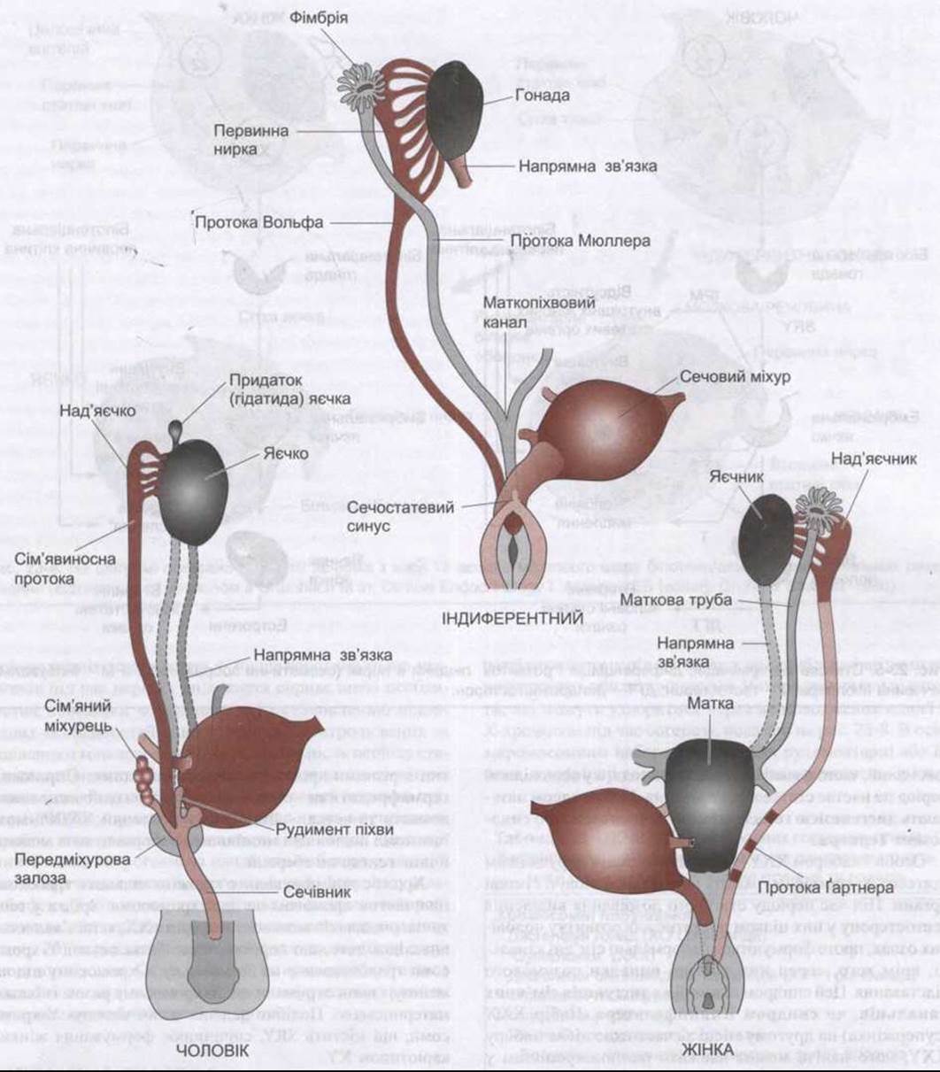
Fig. 23-6. Embryonic differentiation of male and female internal reproductive Organs (genital tracts) from the male (Wolffian) and female (Müllerian) ducts (reproduced with permission from Grumbach M, Conte FA in: Williams Textbook of Endocrinology, 7th ed. Wilson JD, Foster DW [editors], Saunders, 1985).
Hormonal Disorders
Normally, External Male Genitalia develop in genetic males due to the action of androgens secreted by embryonic testes. However, male-type sexual development can also occur in genetic females under METABOLISM/18.html">The Influence of androgens from other sources between 8 and 13 weeks of gestation. The resulting syndrome is called female pseudohermaphroditism. A pseudohermaphrodite is an individual with the genetic constitution and gonads of one sex and the genitalia of the other. It is known that after the 13th week, the genitalia are fully formed, but exposure to androgens can cause clitoral hypertrophy. Female pseudohermaphroditism can be caused both by congenital adrenal hyperplasia with virilization (see Chapter 20) and by maternal administration of androgens. Conversely, the development of female genitalia in genetic males (male pseudohermaphroditism) occurs in the event of embryonic testicular deficiency. Since the testes also secrete MIF, genetic males with deficient testes possess Internal Female Genitalia.
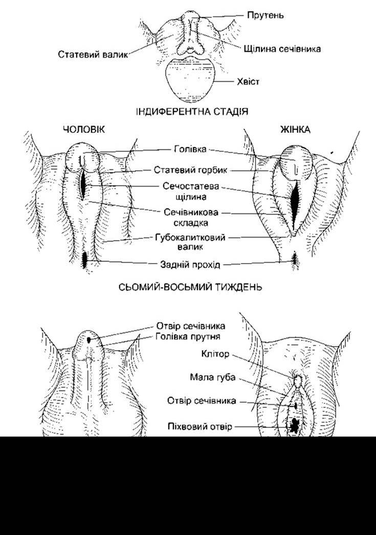
Fig. 23-7. Differentiation of male and female external genitalia from indifferent embryonic structures.
Another cause of male pseudohermaphroditism is androgen resistance, in which male Hormones fail to exert their full effects on Tissues due to various congenital defects. One form of androgen resistance is 5α-reductase deficiency, which results in decreased levels of the enzyme responsible for The production of dihydrotestosterone—the active form of testosterone. The consequences of this deficiency are discussed in the section on the Male Reproductive System. Other forms of androgen resistance are caused by various Mutations in the androgen receptor gene, with receptor dysfunction ranging from mild to severe. Mild impairments lead to Infertility with or without gynecomastia (see below). If the loss of receptor function is substantial, testicular feminization syndrome occurs. In this condition, MIF is present, and testosterone secretion is normal or even enhanced. The external genitalia are female, but the Vagina ends in a blind pouch because internal female genitalia are absent. Individuals with this syndrome develop enlarged breasts during puberty and are considered normal females until evaluated for primary Amenorrhea.
It should be noted that genetic males with a congenital block in pregnenolone synthesis are hermaphrodites, since both testicular and adrenal androgens are normally synthesized from pregnenolone. Male pseudohermaphroditism also occurs in congenital 17α-hydroxylase deficiency (see Chapter 20).

Fig. 23-8. Brief Overview of four possible disorders resulting from maternal sex chromosome nondisjunction during meiosis. The YO chromosomal complement is considered lethal and causes intrauterine fetal death.
Last update: 10/08/2026
Editorial and Educational Adaptation: This material has been compiled based on the primary/original source text. The project team performed an editorial review, corrected technical inaccuracies, structured sections, and adapted the content for an educational format.
What was processed:
- elimination of formatting defects (OCR errors, structural breaks, corrupted characters);
- editorial organization of content;
- standardization of terminology in accordance with academic sources;
- verification of factual statements against the original source text.
All mentions of the author, publication year, and origin of the primary text have been preserved in accordance with the source.