Orthopedics - Oleksa A.P. 2006
Neurogenic arthropathies
Syringomyelic arthropathy
Neurogenic arthropathies are rare and develop As a result of syringomyelia or Syphilis.
Syringomyelic arthropathies are uncommon due to the rarity of the underlying condition, yet they occur more frequently than tabetic arthropathies.
Clinical observations indicate that syringomyelic arthropathies most commonly affect the elbow and hand regions due to cervical Spinal Cord involvement, although lesions in other spinal cord segments and syringomyelic arthropathies of other joints may occur simultaneously with deformities of the Thorax, dorsal spine, and other areas. Thévenard described an ulceromutilating arthropathy with destruction and lysis of the Metatarsal Bones resulting from syringomyelia of the lumbosacral spinal cord.
Syringomyelic arthropathy develops very slowly, progressing gradually. Associated with syringomyelic disorders, thermoanalgesia appears, eventually followed by amyotrophy and sensory disturbances. Patients lose the sensation of heat and frequently burn their fingers. Deformities of the fingers or the elbow develop, which are painless and therefore do not cause the patient any concern.
Upon examination (Fig. 361), the joint is swollen, and the Skin is of normal color and thinned. Local Temperature is also normal. Palpation reveals fluctuation caused by joint effusion. Puncture yields viscous synovial fluid, sometimes mixed with Blood. The range of motion in the joint is fully preserved, and the patient experiences no pain whatsoever. With osteolysis of the articular bone ends, loose bodies appear in the joint, which may become trapped, and the range of painless motion can exceed normal limits. Subluxations or pathological dislocations occasionally occur in the affected joint.
Plain radiographs early in the disease show no pathological changes other than Osteoporosis of the bone metaepiphyses of the joint.
When osteolysis of the articular ends occurs, radiographs reveal distinct destruction with irregular contours and loss of normal anatomical joint configurations, along with the presence of free-floating bone sequestra within the joint (similar to chondromatosis); subluxations and dislocations of the peripheral bone segment may also be observed (Fig. 362).
Blood and urine tests are generally within normal limits.
Patients should receive neurological Treatment for syringomyelia. In cases of joint instability and excessive range of motion, patients are advised to use an orthopedic brace, which enhances limb function and prevents pathological subluxation and dislocation.
Class="center">
Fig. 361. Radiograph of the shoulder joint in a patient with syringomyelia.
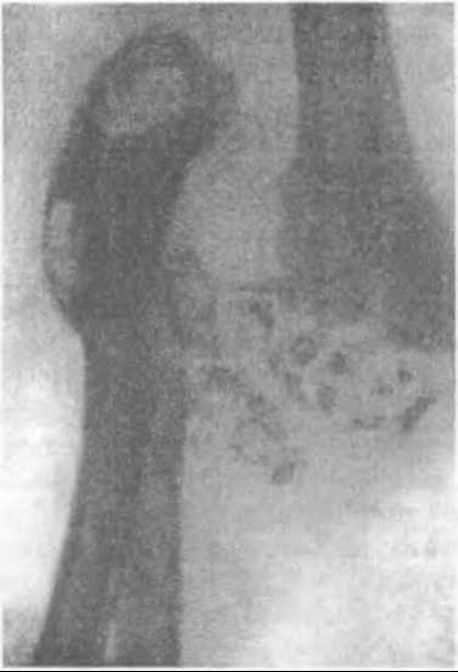
Fig. 362. Radiograph of the elbow joint showing loose bodies and dislocation of the forearm bones due to syringomyelic arthropathy.
Last update: 10/08/2026
Editorial and Educational Adaptation: This material has been compiled based on the primary/original source text. The project team performed an editorial review, corrected technical inaccuracies, structured sections, and adapted the content for an educational format.
What was processed:
- elimination of formatting defects (OCR errors, structural breaks, corrupted characters);
- editorial organization of content;
- standardization of terminology in accordance with academic sources;
- verification of factual statements against the original source text.
All mentions of the author, publication year, and origin of the primary text have been preserved in accordance with the source.