Orthopedics - Oleksa A.P. 2006
Congenital and Acquired Deformities of the Lower Extremity
Lower Extremity Axis Malalignment
Blount's Disease
Blount's disease (Blount-Erlacher-Biesiezny-Barber syndrome) is a localized epiphyseal Dysplasia affecting the proximal tibial epiphysis. It involves multiplanar deformity of the Tibia with a predominantly varus angulation of the metaepiphyseal region, which progressively worsens as the child grows (Fig. 190). This leads to secondary static deformities in the ankle joint and the FOOT.
Uneven weight-bearing distribution across the tibial and femoral physis results in knee joint instability. This is caused by overstretching of the collateral ligament on the convex side of the knee and stress concentration zones in localized areas of the articular Cartilage, ultimately triggering early-onset degenerative joint changes (gonarthrosis) during childhood.
Parents typically seek medical attention due to unilateral or bilateral varus knee deformity and a limp. The children themselves are painless.
Physical examination reveals varus bowing of the shin around the knee, or bow-legs (O-shaped lower limbs) in bilateral cases. Range of motion in the knee is full and pain-free. Testing for lateral knee laxity with the leg fully extended may reveal insufficiency of the collateral ligament opposite the concavity of the tibial curvature. Joint effusion is generally absent. In adolescent and older patients, crepitus may be felt during joint movement, indicating structural damage to the articular cartilage.
Radiographs of the medial portion of the tibial growth plate reveal narrowing and an irregular, wavy appearance compared to the lateral side (Fig. 191). Sclerosis of the Bone tissue adjacent to the growth plate is often present. The joint space is narrowed on the affected side and widened on the contralateral side. The tibia shows varus bowing at the level of the proximal metaphysis.
Class="center">
Fig. 190. Varus deformity of both tibiae in Blount's disease.
Routine Blood and urine laboratory values are within normal limits.
Diagnosing Blount's disease is generally straightforward.
Treatment. Conservative management is indicated for children aged 3 to 4 years. Custom corrective braces are prescribed for daily wear and adjusted as the child grows. Nighttime treatment involves bandaging a straight splint to the lateral aspect of the lower limb. Muscle massage and physical therapy are also recommended.
To stimulate epiphyseal cartilage growth, a bone peg or allograft is sometimes inserted into a drilled channel beneath the physis, though clinical studies have not confirmed the efficacy of this method.
Conservative treatment helps prevent the rapid progression of the varus deformity, but it does not correct it.
Surgical intervention is indicated for tibial varus deformity exceeding 20°. Surgery is performed under general anesthesia with a tourniquet. Dissection is carried out down to the bone. Subperiosteal corrective osteotomy is performed either below or above the tibial tuberosity, distal to the growth plate, and the limb axis is realigned. If acute correction is successfully achieved, the bone fragments are stabilized with one or two percutaneous Kirschner wires, followed by the application of a hip spica cast.
If achieving a normal limb axis intraoperatively is unsuccessful, gradual correction is carried out in the postoperative period.
In adolescents, acute correction of a severe varus deformity is difficult; therefore, the Procedure is supplemented by a fibular osteotomy and completed with the application of an Ilizarov fixator, which allows for the gradual restoration of the lower limb axis (Fig. 192).
It is worth noting that surgery is best performed after major growth spurts to prevent recurrence. Parents must be warned about the potential recurrence of the deformity following subsequent periods of rapid growth. Therefore, in 4- to 5-year-old children, the tibia is slightly overcorrected during osteotomy into a mild valgus alignment, as natural growth will eventually straighten the limb.
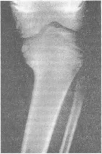
Fig. 191. Radiograph of the tibia in Blount's disease.

Fig. 192. Metaphyseal osteotomy of the tibia and slightly distal Fibula (n.peroneus) following closure of the growth plate in Blount's disease.
Last update: 10/08/2026
Editorial and Educational Adaptation: This material has been compiled based on the primary/original source text. The project team performed an editorial review, corrected technical inaccuracies, structured sections, and adapted the content for an educational format.
What was processed:
- elimination of formatting defects (OCR errors, structural breaks, corrupted characters);
- editorial organization of content;
- standardization of terminology in accordance with academic sources;
- verification of factual statements against the original source text.
All mentions of the author, publication year, and origin of the primary text have been preserved in accordance with the source.