Orthopedics - Oleksa A.P. 2006
Examination of the orthopedic patient
Assessment of joint range of motion
A normal joint is characterized by its regular shape, pain-free and full-range motion, and well-developed Muscles that facilitate these movements.
Joint range of motion is the primary functional indicator, and its limitation is the main reason a patient seeks medical attention. The range of motion is measured with a goniometer and recorded in degrees. The arms of the goniometer are placed along the axis of the two limb segments connected by the joint, with the fulcrum of the goniometer corresponding to the axis of motion in the joint (Fig. 16).
Class="center">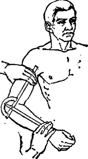
Fig. 16. Measuring the range of motion in the elbow joint using a goniometer.
Active movements are performed independently by the patient, whereas passive movements are carried out by external force. In each joint, the direction and range of motion vary depending on its anatomical Structure and Functional purpose. It is worth noting that in children and athletes, the amplitude of joint motion is slightly greater than the physiological norm for an adult.
In the frontal plane, adduction (adductio) or abduction (abductio) of a limb segment occurs, while in the sagittal plane, flexion (flexio) or extension (extensio) takes place. Movements around the longitudinal axis of a limb or its segment are called rotation (rotacio). Lateral rotation (supinatio) and medial rotation (pronatio) of the forearm are distinguished.
The degree of limb movement variation in different planes is measured from its initial position, that is, the position the limb assumes when The Human Body is in a relaxed vertical posture. For both upper and lower limbs, the neutral starting position is considered 180°, which in Western European countries is taken as 0°. Measuring the amplitude of movements helps determine the degree of deviation from the norm as well as the effectiveness of Treatment.
During a patient examination, the amplitude of active movements should be assessed first, followed by passive movements. When evaluating motion in the shoulder joint, the scapula must be fixed, because the functional capability of the upper limb relies not only on movement within the shoulder joint itself, but also on the Displacement of the shoulder girdle in various planes. One arm of the goniometer is placed along the body axis, and the other along the axis of the arm. Normally, shoulder abduction is possible up to 90°, flexion up to 130–135°, and extension up to 35–45°. Rotational Movements of the shoulder around its axis occur both medially and laterally within a range of 20–35° (Fig. 17). In the elbow joint, the total range of motion is 150–155°, allowing for full extension (from 180°) and maximum flexion (up to 25–30°). In children and women, Normal Anatomy may allow for additional forearm hyperextension of up to 5° (Fig. 18).
In the wrist joint, palmar flexion of the hand (toward the palmar surface) is possible up to 50–75°, extension up to 50–60°, radial deviation (toward the radial side) up to 20–30°, and ulnar deviation (toward the ulnar side) up to 35–40°. The amplitude of pronation-supination movements ranges between 80–90° (Fig. 19).
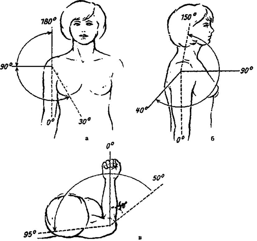
Fig. 17. Determining the amplitude of motion in the shoulder joint: a — shoulder adduction and abduction, b — flexion and extension, c — rotational movements of the shoulder.
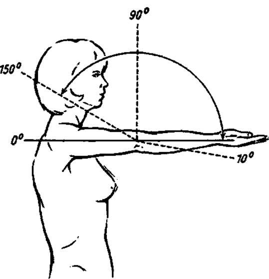
Fig. 18. Range of motion in the elbow joint.
When determining the degree of flexion, extension, adduction, and abduction of the hip, it should be kept in mind that these movements can be produced not only by the hip joint itself, but also compensated by the spine through an increase or decrease in lumbar lordosis and lateral movements. Therefore, in cases of flexion-adduction contractures of the hip joint, to eliminate lordosis, it is advisable to fix the pelvis and the lumbar spine by maximum flexion (toward the abdomen) of the contralateral thigh so that the linea bispinalis remains perpendicular to the body axis. Normally, the range of motion of the hip joint in the sagittal plane is 120–140° and is achieved primarily through hip flexion. Additional hip extension (hyperextension) is possible by 10–15°. In the frontal plane, hip abduction ranges from 30–50°, and adduction from 35–45°. Rotational movements of the hip in the joint are possible by 45° both inward and outward. Medial and lateral Rotation of the hip are determined by the deviation in either direction of the lower leg flexed at 90° in the knee joint (Figs. 20, 21).

Fig. 19. Normal adduction and abduction of the hand, along with its flexion and extension.
Movements in the knee joint occur primarily in the sagittal plane — flexion ranges from 180° to 45–50°. Normally, a slight degree of knee hyperextension (by 5–10°) is also possible (Fig. 22).
In the ankle joint, plantarflexion ranges from 45–60°, and dorsiflexion from 25–35° (Fig. 23). In the equinus FOOT position, movements in the ankle and small tarsal joints allow for supination and pronation of the foot within 45–55°, as well as adduction and abduction within 30° (Fig. 24).
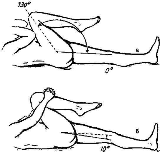
Fig. 20. Determining the neutral position of the limb at the hip joint: a — at maximum passive flexion, b — while the contralateral thigh is flexed by 10°.

Fig. 21. Determining normal angles: a — hip adduction and abduction, b — its rotational movements.
Spinal movements are multiaxial and are performed by tilting or rotating the HEAD AND TORSO. In the cervical spine, flexion (bending the head forward) is 65–75°, and extension is 40–50°. Rotational movements of the head in both directions are possible by 40–45° (Fig. 25). Forward trunk flexion is 75–90°, and backward extension is 35–40°. Lateral trunk movements occur primarily due to the lumbar spine and measure 35–40°, while rotational movements measure 20–30° (Fig. 26).
Deviations from the normal range of motion in a joint can occur in the direction of either an increase or a decrease. An increase in the range of motion beyond physiological limits occurs during ligamentous injuries or overstretching of the ligamentous-capsular apparatus, intra-articular fractures, inflammatory joint processes, after poliomyelitis, etc. In such cases, deformation or subluxation of the articular bone end typically occurs, accompanied by abnormal laxity and joint instability (by fixing the proximal limb segment with one hand, the examiner assesses the degree of pathological mobility of the peripheral segment with the other).
Limitation of joint mobility varies in both duration and scope. It can be temporary and transient, or persistent and chronic.

Fig. 22. Range of normal knee flexion (150°) and additional hyperextension (10°).
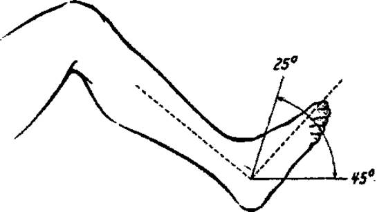
Fig. 23. Normal range of foot flexion and extension.
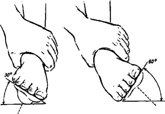
Fig. 24. Assessment of normal foot pronation and supination.
An example of temporary movement restriction and initial pain is deforming osteoarthritis of the knee joints, where pain leads to Muscle tension. All of this disappears once the patient "walks it off." A specific type of morning stiffness can be observed in patients with rheumatoid Arthritis. They find it difficult to move after waking up, but after a few hours, the stiffness subsides and the range of motion in the joints increases or returns to normal. This phenomenon is associated with hypocortisolism, increased synovial fluid viscosity, and other factors.

Fig. 25. Normal range of motion in the cervical spine: a - forward and backward head flexion, b - lateral head bending, c - head rotation.
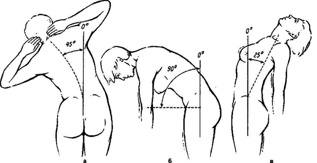
Fig. 26. Assessment of normal spinal range of motion: a - lateral bending, b - during flexion, c - during extension.
However, cases of persistent limitation of joint movement are frequently encountered, which, regardless of the cause, is referred to as joint contracture.
If joint movement is sharply restricted and only minimal movements (within 3-5°) can be detected, this condition is termed joint rigidity. It typically results from previous purulent adhesive arthritis or similar conditions.
Complete absence of movement in a joint is called ankylosis. Furthermore, ankylosis can be fibrous (ancylosis fibrosa) or bony (ancylosis ossea) (Fig. 27). They can be differentiated only by radiography. If there is no movement in the joint, but the joint space is preserved, this indicates fibrous ankylosis, whereas if the space is obliterated by Bone tissue, it is bony ankylosis. Ankylosis, much like joint contracture, can occur in either a functionally favorable or functionally unfavorable position of the limb.
If malpositions occur in such a way that the patient can perform vital Functions with extreme effort using their limbs (eating, dressing, walking, etc.), they are termed concordant. Limb deformities that prevent the patient from performing these activities are called discordant.
Discordant deformities of the upper and lower limbs are quite common in patients with rheumatoid arthritis or spastic paralysis. For example, in the presence of severe (acute-angle) flexion contractures of both knee joints and flexion-adduction contractures of the hip joints, the patient is unable to move even with the aid of crutches.
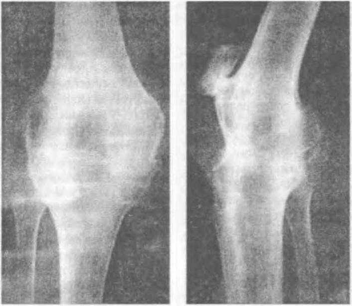
Fig. 27. Bony ankylosis of the knee joint: radiographs in two projections.
Last update: 10/08/2026
Editorial and Educational Adaptation: This material has been compiled based on the primary/original source text. The project team performed an editorial review, corrected technical inaccuracies, structured sections, and adapted the content for an educational format.
What was processed:
- elimination of formatting defects (OCR errors, structural breaks, corrupted characters);
- editorial organization of content;
- standardization of terminology in accordance with academic sources;
- verification of factual statements against the original source text.
All mentions of the author, publication year, and origin of the primary text have been preserved in accordance with the source.