Special Histology and Embryology: Practical Course - V. K. Naphanyuk 2001
Digestive System
Anterior Segment of the Digestive System
Esophagus
The Esophagus is a muscular tube approximately 30 cm in length that connects the Pharynx to The Stomach.
The esophageal wall consists of four layers: the mucosa, submucosa, muscular layer, and adventitia.
The mucosal layer is composed of the epithelium, lamina propria, and muscularis mucosae.
The epithelium of the mucosa is stratified squamous non-keratinized. The epithelial layer consists of 20-25 Cell layers.
The lamina propria forms Connective Tissue papillae that project into the epithelium. It contains two groups of glands (esophageal cardiac glands). One group is located at the level of the laryngeal cricoid Cartilage and the fifth tracheal ring. The second group is situated near the entrance to the stomach. These are simple branched tubular glands that produce a mucous secretion. In addition, the terminal portions of the cardiac glands contain numerous endocrine Cells.
The muscularis mucosae of the esophagus consists of longitudinally arranged bundles of smooth Muscle cells surrounded by a network of elastic fibers.
The submucosa is formed by loose connective tissue housing the terminal secretory portions of the esophageal proper glands. These are complex, heavily branched tubuloalveolar glands with a mucous type of secretion. As a rule, they are located on the ventral surface of the upper third of the esophagus and consist exclusively of mucocytes.
The muscular layer of the esophagus consists of an inner circular and an outer longitudinal layer, separated by layers of loose connective tissue.
In the upper third of the esophagus, these layers are composed of Skeletal Muscle tissue. In the middle third, they consist of both skeletal and Cytology/cytology/32.html">Smooth Muscle tissue.
In the lower third of the esophagus, they consist entirely of smooth muscle tissue.
The adventitia is connected on one side with the connective tissue septa located within the muscular layer, and on the other side with the surrounding connective tissue of the Mediastinum. The abdominal segment of the esophagus is covered by a serous membrane formed by mesothelium with underlying connective tissue.
Slide 13. Longitudinal section of the esophagus (Fig. 49).
Low magnification. Examine and sketch the slide. Identify the layers of the esophageal wall.
The mucosa consists of stratified squamous epithelium and the lamina propria, which projects into the epithelium in the form of papillae. Numerous cross-sections of small Blood Vessels can be seen in this layer. This is followed by the muscularis mucosae, formed by loosely arranged bundles of longitudinal smooth muscle fibers, which appear sectioned longitudinally on the slide. The submucosa is formed by loose connective tissue containing nerve plexuses and large blood vessels. Clusters of terminal glandular units are identified in this layer. Their mucous nature is indicated by the foamy basophilic secretion.
Class="center">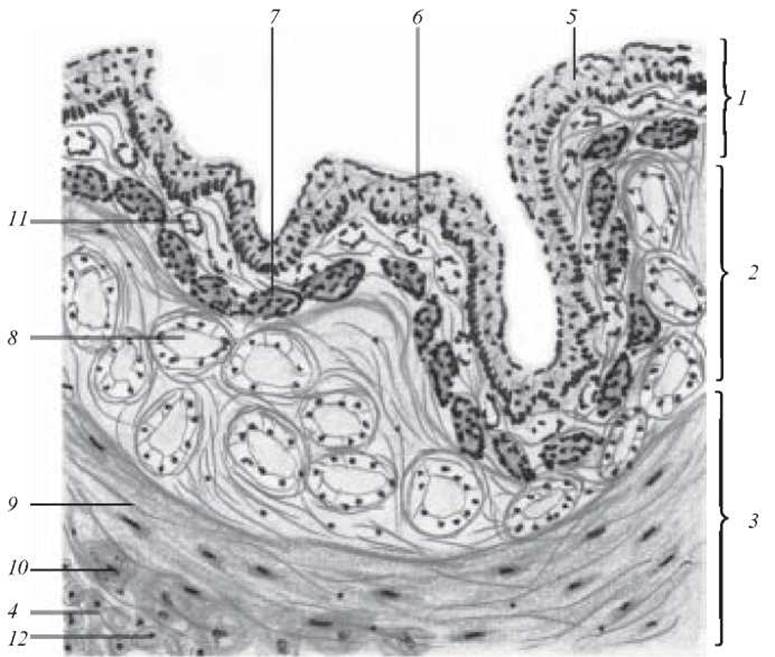
Fig. 49. Cross-section of the esophagus. Hematoxylin-eosin staining. x 160:
1 — mucosa; 2 — submucosa; 3 — muscular layer; 4 — adventitia; 5 — stratified squamous epithelium of the mucosa; 6 — lamina propria of the mucosa; 7 — muscularis mucosae; 8 — glands; 9 — circular layer of the muscular coat; 10 — longitudinal layer of the muscular coat; 11 — blood vessels; 12 — fat cells
The muscular layer consists of an inner circular and an outer longitudinal layer, between which lies the myenteric (Auerbach's) nerve plexus.
The adventitia covers the external surface of the esophagus (from the mediastinal side). It is loose irregular connective tissue containing blood vessels, adipose tissue, nerve bundles, and the adventitial nerve plexus.
On the drawing, label: 1) mucosa; 2) submucosa; 3) muscular layer; 4) adventitia; 5) stratified squamous epithelium of the mucosa; 6) lamina propria of the mucosa; 7) muscularis mucosae; 8) glands; 9) excretory duct of the gland; 10) circular layer of the muscular coat; 11) longitudinal layer of the muscular coat; 12) blood vessels; 13) fat cells.
Slide 14. Cross-section of the esophagus at the gastroesophageal junction (Fig. 50).
Low magnification. Locate the transition zone. At this site, there is an abrupt transition from the stratified squamous epithelium of the esophagus to the simple columnar epithelium of the stomach, which forms gastric pits. Cardiac glands are located in the lamina propria of both the esophagus and the stomach. These are simple branched tubular glands composed of the following cell types: chief cells (exocrinocyti principales), parietal cells (exocrinocyti parietales), and endocrine cells (endocrinocyti gastrointestinales). The muscularis mucosae of the esophagus continues into the stomach.
The esophageal submucosa contains proper esophageal glands. They are numerous in this preparation and occasionally extend into the submucosa of the stomach.
The muscular layer consists of two layers: an inner circular layer, which forms a sphincter, and an outer longitudinal layer.
The subdiaphragmatic region of the esophagus (abdominal part) and the cardiac region of the stomach are externally covered by a serous membrane.
On the figure, indicate: 1) gastric columnar epithelium; 2) gastric pit; 3) cardiac gland; 4) stratified squamous epithelium of the esophagus; 5) lamina propria of the mucosa; 6) muscularis mucosae; 7) submucosa; 8) muscular layer; 9) serosa.
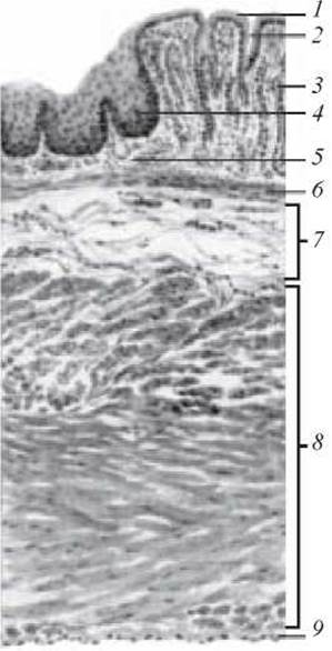
Fig. 50. Cross-section of the esophagus at the gastroesophageal junction. Hematoxylin and eosin staining. x 40:
1 — gastric columnar epithelium; 2 — gastric pit; 3 — cardiac gland; 4 — stratified squamous epithelium of the esophagus; 5 — lamina propria of the mucosa; 6 — muscularis mucosae; 7 — submucosa; 8 — muscular layer; 9 — serosa
Last update: 10/08/2026
Editorial and Educational Adaptation: This material has been compiled based on the primary/original source text. The project team performed an editorial review, corrected technical inaccuracies, structured sections, and adapted the content for an educational format.
What was processed:
- elimination of formatting defects (OCR errors, structural breaks, corrupted characters);
- editorial organization of content;
- standardization of terminology in accordance with academic sources;
- verification of factual statements against the original source text.
All mentions of the author, publication year, and origin of the primary text have been preserved in accordance with the source.