Medical Radiology - Lazar A.P. 2008
Radiation examination of bones and joints
Traumatic injuries of bones and joints
Radiological examination is indicated for all injuries of The Musculoskeletal System to detect fractures, dislocations, subluxations, and foreign bodies, as well as to monitor fragment reposition, the appearance of bone callus, etc. X-ray imaging remains the primary diagnostic method, while sonography, CT, and MRI are performed according to specific clinical indications. Ultrasound and MRI are particularly effective in identifying soft tissue INJURIES OF THE musculoskeletal system, including tears and strains of Muscles, tendons, ligaments, and the Joint Capsule, as well as post-traumatic fluid accumulation (Fig. 40). Non-ionizing imaging Methods are also especially valuable for newborns and children. CT provides superior visualization of cranial and pelvic bone fractures and fissures, foreign bodies, and surrounding soft tissue changes that often remain undetected on standard radiographs.
Class="center">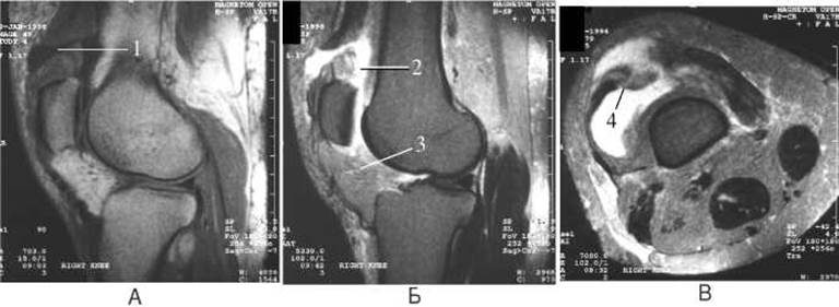
Fig. 40. Quadriceps tendon rupture on Magnetic Resonance imaging: sagittal T1-weighted (A) and T2-weighted (B), and axial T2-weighted (C) images. The images demonstrate tendon rupture (1), free fluid surrounding the rupture site (2), patellar tendon deformation (3), and lateral patellar subluxation (4).
Radiographically, bone fractures are identified by the following signs: a fracture line, fragment displacement, disruption of Bone Structure, and cortical bone layer interruption (Fig. 41). The fracture line typically appears as a radiolucent band with sharp margins. This radiolucent band is most commonly straight and less frequently jagged. The bone trabecular pattern of the adjacent fragments remains unchanged. In impacted fractures, the fracture line appears as a ribbon-like shadow against the Background of the normal surrounding bone pattern.
If the fracture line extends across the entire width of the bone, it is classified as a complete fracture; if it extends across more than half of the width, it is termed a fissure; and if less than half, it is considered an infraction (partial fracture). Based On the Relationship between the fracture line and the joint, fractures are categorized as intra-articular, peri-articular, or extra-articular.
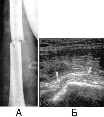
Fig. 41. Humeral fracture on a radiograph (A) and sonogram (B).
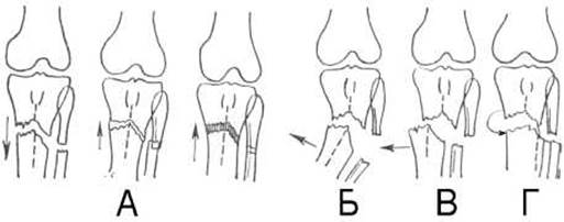
Fig. 42. Types of bone fragment displacement:
A - along the length;
B - at an angle;
C - along the width;
D - with rotation.
Disruption of the cortical layer is evident even when the fracture line and fragment displacement are not radiologically apparent, such as in subperiosteal fractures in children. Alteration of the bone structure in fractures is detected by examining the alignment and trajectory of bone trabeculae. Indirect radiological signs of a fracture include bone deformation, small bone fragments in the soft Tissues, a hematoma shadow, and several others.
Bone fragment displacements are classified as width displacement (lateral), length displacement (with overriding, Separation, or impaction), axial displacement (angular), peripheral displacement (involving rotational turning of the fragment), and combined displacement, which involves multiple types of shifting simultaneously (Fig. 42).
A dislocation is defined as a complete disruption of the congruency of the articular surfaces of bones (Fig. 43), whereas a subluxation represents an incomplete disruption of this congruency. Subluxation is typically characterized by a wedge-shaped radiographic joint space and axial deviation of the dislocated bone.
In children, fractures are largely determined by the unique structural properties of their bones, which are thinner and more flexible. Their periosteum is elastic and tends to maintain its integrity during various traumas more often than the underlying bone trabeculae. Radiologically, bone displacement and the radiolucent band may be absent; however, a cortical step-off deformity is visible at the fracture site along the outer edge of one or both bone contours at the level of trauma. Such injuries are known as subperiosteal fractures, or "greenstick" fractures (Fig. 44).

Fig. 43. Dislocation of the lower leg on a lateral radiograph of the knee joint.

Fig. 44. Greenstick fracture of the distal radius on a radiograph (diagram).
Another distinct category of pediatric injuries involves fractures occurring within the growth plate, which is composed of cartilaginous tissue. These fractures are frequently accompanied by Displacement of the ossification center and are referred to as epiphyseolyses (Figs. 45, 46).
In elderly and senile individuals, bone fragility causes fractures to occur even from minor trauma. These are typically comminuted, featuring significant fragment displacement. Such fractures are most frequently localized in the femoral and humeral necks, as well as the distal metaphyses of the radius and leg bones. Fractures in older adults heal 2 to 3 times slower than in younger individuals.
During the fracture healing process, the first 7–10 days involve the resorption of damaged bone trabeculae and The formation of a primary Connective Tissue callus; radiographs during this phase reveal a widening of the fracture line. Over the subsequent 7–10 days, the connective tissue callus transforms into osteoid tissue devoid of calcium salts, which is why radiological changes are typically absent during this period. After the 20th day, a true bony callus forms, which is clearly visible on radiographs by days 30–35.
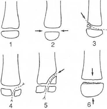
Fig. 45. Types of bone end fractures in children on radiographs (diagram).
1 - normal;
2 - epiphysiolysis with widening of the radiolucent zone (9% of cases);
3 - osteoepiphysiolysis with metaphyseal fracture (70%);
4 - osteoepiphysiolysis with epiphyseal fracture (10%);
5 - metaepiphyseal fracture involving the epiphyseal plate (10%);
6 - compression of the epiphyseal Cartilage along the limb axis, not visible on initial radiographs (1%).

Fig. 46. Osteoepiphysiolysis of the medial malleolus on an anteroposterior radiograph of the ankle region.
The indicated timeframes for callus formation depend on age, the anatomical site of the injury, and individual patient characteristics.
When fracture healing is delayed due to inadequate reduction and immobilization of the fragments, or soft tissue interposition between them, a pseudarthrosis (false joint) develops at the fracture site with the following radiographic features: 1) a distinct radiolucent fracture line is visible; 2) no callus formation is detected despite sufficient time having elapsed; 3) the ends of the fragments appear somewhat rounded and smoothed; 4) the medullary cavity becomes sealed by a compact bone plate; 5) a socket forms on one fragment, while the end of the other fragment becomes rounded. Later, the radiolucent line between the fragments widens as the newly formed articulating surfaces of the socket and HEAD become covered with cartilage.
If fractures occur as a complication of underlying bone diseases (tumors, metastases, cysts, Osteomyelitis), they are termed pathological fractures and are characterized by an altered bone structure in the fracture area.
Spinal trauma frequently involves fractures of the vertebral bodies. Such fractures are referred to as compression fractures. They are accompanied by a reduction in the height of the vertebral body and its wedge-shaped deformity (Fig. 47).
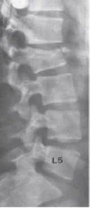
Fig. 47. Fracture of the body of the second lumbar vertebra on a lateral radiograph of the lumbar spine.
Last update: 08/08/2026
Editorial and Educational Adaptation: This material has been compiled based on the primary/original source text. The project team performed an editorial review, corrected technical inaccuracies, structured sections, and adapted the content for an educational format.
What was processed:
- elimination of formatting defects (OCR errors, structural breaks, corrupted characters);
- editorial organization of content;
- standardization of terminology in accordance with academic sources;
- verification of factual statements against the original source text.
All mentions of the author, publication year, and origin of the primary text have been preserved in accordance with the source.