Human Anatomy - Kotsan I. Y. 2009
Skeleton of the lower limb
Bones of the leg
The crus (leg) consists of two bones: the Tibia and the Fibula (Fig. 71).
Class="center">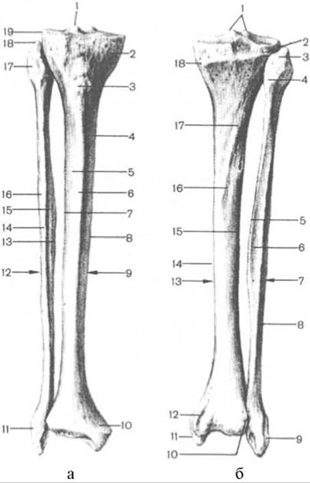
Fig. 71. Tibia and fibula (right bones)
a — anterior view: 1 — intercondylar eminence, 2 — medial condyle, 3 — tibial tuberosity, 4 — medial surface, 5 — lateral surface, 6 — anterior border, 7 — interosseous border, 8 — medial border, 9 — tibia, 10 — medial malleolus, 11 — lateral malleolus, 12 — fibula, 13 — interosseous border, 14 — anterior border, 15 — medial surface, 16 — lateral surface, 17 — HEAD of fibula, 18 — apex of fibular head, 19 — lateral condyle of tibia, b — posterior view: 1 — lateral and medial intercondylar tubercles, 2 — lateral condyle, 3 — apex of fibular head, 4 — head of fibula, 5 — interosseous border, 6 — posterior surface, 7 — fibula, 8 — lateral surface, 9 — lateral malleolus, 10 — articular surface of malleolus, 11 — medial malleolus, 12 — malleolar sulcus, 13 — tibia, 14 — medial border, 15 — interosseous border, 16 — posterior surface, 17 — soleal line, 18 — medial condyle
The tibia is located on the medial side of the leg. It is a long tubular bone consisting of a shaft (body) and two ends: proximal and distal.
The proximal end of the tibia is significantly thickened and expanded, featuring two condyles: medial and lateral (condylus medialis et condylus lateralis). On the superior aspect of the condyles lies the superior articular surface (facies articularis superior), which articulates with the condyles of the Femur. The superior articular surface of the condyles is divided in the middle by the intercondylar eminence (eminentia intercondylaris), which consists of the medial intercondylar tubercle (tuberculum intercondylare mediale) and the lateral intercondylar tubercle (tuberculum intercondylare laterale). Anterior to the intercondylar eminence is the anterior intercondylar area (area intercondylaris anterior), and posteriorly lies the posterior intercondylar area (area intercondylaris posterior). On the inferolateral aspect of the lateral condyle, somewhat posteriorly, is the flat fibular articular surface (facies articularis fibularis), which serves for articulation with the fibula.
The shaft of the tibia (corpus tibiae) is triangular in shape. It presents three surfaces: medial, lateral, and posterior. These are separated from one another by three borders: anterior, medial, and lateral (interosseous).
The medial surface (facies medialis) is smooth and lies directly beneath the Skin. The lateral surface (facies lateralis) is slightly concave and covered by Muscles. The posterior surface (facies posterior) is flat and bears the rough soleal line (linea musculi solei), which runs obliquely downward and medially from the posterior border of the lateral condyle. Below this line is the nutrient foramen (foramen nutricium), which continues into a distally directed nutrient canal (canalis nutricius).
The anterior border (margo anterior) of the tibia is sharp and easily palpable through the skin. In the upper part of the bone, it thickens to form the tibial tuberosity (tuberositas tibiae), to which the quadriceps femoris Muscle attaches. The medial border (margo medialis) is slightly rounded. The lateral, or interosseous, border (margo interosseus) is sharp and faces the corresponding border of the fibula. The interosseous membrane of the leg attaches to it.
The distal end of the tibia is expanded and features a downward projection on its medial side—the medial malleolus (malleolus medialis)—and the fibular notch (incisura fibularis) on its lateral side for articulation with the fibula. Behind the medial malleolus runs the shallow malleolar sulcus (sulcus malleolaris), which lodges the tendon of the tibialis posterior muscle. On the lateral side of the medial malleolus is the articular surface of the malleolus (facies articularis malleoli), which transitions at an angle into the inferior articular surface (facies articularis inferior). Together with the articular surface of the fibula, these surfaces articulate with the talus bone of the FOOT.
The fibula (fibula) is a slender, long tubular bone located on the lateral side of the leg. It has a shaft and two ends: proximal and distal.
The proximal end of the bone bears the head of the fibula (caput fibulae). Superiorly, the head terminates in a pointed apex of the fibular head (apex capitis fibulae). Inferiorly, the head gradually tapers into the neck of the fibula (collum fibulae). On the medial side of the head is the articular surface of the fibular head (facies articularis capitis fibulaе), which serves for articulation with the tibia.
The shaft of the fibula (corpus fibulae) resembles a triangular prism slightly twisted around its longitudinal axis. The shaft presents three surfaces: lateral, medial, and posterior (facies lateralis, facies medialis et facies posterior), which are separated from one another by three borders: anterior, posterior, and interosseous (margo anterior, margo posterior et margo interosseus).
The distal end of the fibula thickens to form the lateral malleolus (malleolus lateralis). On the Medial surface of the lateral malleolus is the smooth articular surface of the malleolus (facies articularis malleoli). Posterior to the articular surface of the malleolus lies the malleolar fossa of the lateral malleolus (fossa malleoli lateralis), which is the site of attachment for the posterior talofibular ligament. On the posterior aspect of the lateral malleolus, a flat malleolar sulcus (sulcus malleolaris) is visible, marking the passage of the fibular (peroneal) tendons. Throughout almost its entire length, the fibula is covered by a thick layer of muscles, and only its lateral malleolus lies directly subcutaneous and is clearly outlined.
Last update: 08/08/2026
Editorial and Educational Adaptation: This material has been compiled based on the primary/original source text. The project team performed an editorial review, corrected technical inaccuracies, structured sections, and adapted the content for an educational format.
What was processed:
- elimination of formatting defects (OCR errors, structural breaks, corrupted characters);
- editorial organization of content;
- standardization of terminology in accordance with academic sources;
- verification of factual statements against the original source text.
All mentions of the author, publication year, and origin of the primary text have been preserved in accordance with the source.