Human Anatomy and Physiology - N. I. Fedyukovich 2003
Internal Organs
Respiratory System
Trachea and Bronchi
The Trachea (trachea) is an unpaired organ through which air enters the Lungs and vice versa (Fig. 80).
The trachea is a tube-shaped organ 9—10 cm long, slightly flattened anteroposteriorly; its average diameter is 15—18 mm.
The framework of the trachea consists of 16—20 hyaline cartilaginous semi-rings connected to each other by anular ligaments.
The trachea begins at the level of the lower border of the VI cervical vertebra and ends at the level of the upper border of the V thoracic vertebra.
The trachea is divided into cervical and thoracic parts. Anterior to the trachea in its cervical part lies The Thyroid Gland, posterior is the Esophagus, and laterally are the neurovascular bundles (common carotid artery, Internal jugular vein, Vagus nerve).
In the thoracic part, anterior to the trachea are the arch of the aorta, the brachiocephalic trunk, the left brachiocephalic vein, the Water/144.html">Origin of the left common carotid artery, and the Thymus gland.
Class="center">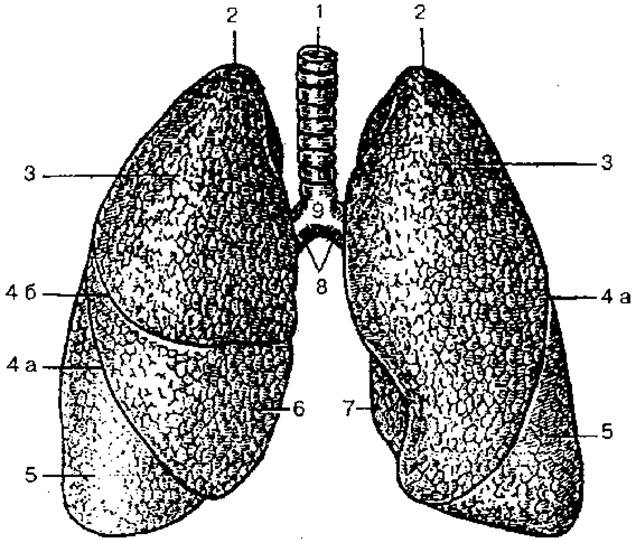
Fig. 80. Trachea, main Bronchi, and lungs:
1 — trachea; 2 — apex of the lung; 3 — superior lobe; 4 a — oblique fissure; 4 b — horizontal fissure; 5 — inferior lobe; 6 — middle lobe; 7 — cardiac notch of the left lung; 8 — main bronchi; 9 — bifurcation of the trachea
In the thoracic cavity, the trachea divides into two main bronchi, which pass to the right and left lungs. The site of tracheal division is called the bifurcation. The right main bronchus has a more vertical course; it is shorter and wider than the left. Consequently, foreign bodies from the trachea more frequently enter the right bronchus. The length of the right bronchus is about 3 cm, and the left is 4—5 cm. The arch of the aorta lies over the left main bronchus, and the azygos vein lies over the right. The right main bronchus has 6—8, and the left has 9—12 cartilaginous semi-rings. Internally, the Trachea and Bronchi are lined with a mucous membrane featuring ciliated pseudostratified epithelium, containing mucous glands and Solitary lymphoid nodules. Externally, the trachea and main bronchus are covered by adventitia.
The main bronchi (primary) in turn divide into lobar (secondary) bronchi, and these in turn divide into segmental (tertiary) bronchi, which branch further to form the bronchial tree of the lungs.
The main bronchi consist of incomplete cartilaginous rings; in medium-sized bronchi, hyaline Cartilage tissue is replaced by elastic cartilage; in terminal bronchioles, the cartilaginous sheath is absent.
Last update: 08/08/2026
Editorial and Educational Adaptation: This material has been compiled based on the primary/original source text. The project team performed an editorial review, corrected technical inaccuracies, structured sections, and adapted the content for an educational format.
What was processed:
- elimination of formatting defects (OCR errors, structural breaks, corrupted characters);
- editorial organization of content;
- standardization of terminology in accordance with academic sources;
- verification of factual statements against the original source text.
All mentions of the author, publication year, and origin of the primary text have been preserved in accordance with the source.