Human Anatomy Part 1 - K. A. Dyubenko, A. K. Kolomiyytsev, Yu. B. Chaykovsky 2002
Special Part
Reproductive systems, systemata genitalia - Female reproductive system, systema genitale femininum
Internal female genital organs, organa genitalia feminina interna - Uterus
The Uterus (uterus s. metra) is a hollow, pear-shaped muscular organ (see Fig. 326, 328) located in the lesser pelvis of a female. It serves for The Development of the embryo in the event of egg Fertilization, as well as for the expulsion of the fetus during childbirth (reproductive function).
The following PARTS OF THE uterus are distinguished: the fundus (fundus uteri), directed upward and forward; the body (corpus uteri), which is the middle triangular part; and the lower part — the cervix (cervix uteri). The lower end of the cervix communicates with the Vagina. The narrowest region where the body of the uterus transitions into the cervix is called the isthmus (isthmus uteri). The portion of the cervix that extends into the vagina is called the vaginal part (portio vaginalis cervicis), while the part outside it is the supravaginal part (portio supravaginalis cervicis).
The uterus has two surfaces: the anterior or vesical surface (facies vesicalis) and the posterior or intestinal surface (facies intestinalis), as well as two borders: the right and left borders (margo uteri dexter et sinister).
The uterine cavity (cavum uteri) is small and triangular in shape (see Fig. 328). The uterine tubes open into it from above and laterally. Below, the cavity transitions into the cervical canal (canalis cervicis uteri), the mucosa of which contains glands (gll. cervicales). The cervical canal opens into the vagina through the uterine orifice (ostium uteri). This opening is bounded by two Lips: the anterior lip (labium anterius) and the posterior lip (labium posterius).
The cervix is conical in young girls, whereas in adult women, especially those who have given birth, it is cylindrical. In nulliparous women, the orifice is transversely oval, while in parous women, it forms a transverse slit (Fig. 329).
Class="center">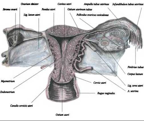
Fig. 328. Cross-section of the uterus (uterus), Uterine tube (tuba uterina), Ovary (ovarium), and vagina (vagina)
The dimensions of the uterus in sexually mature women vary. For instance, in nulliparous women, the length of the uterus is 7-8 cm, whereas in parous women it is 8-9.5 cm; the width at the site of Water/144.html">Origin of the uterine tubes is 4-4.5 cm. The weight of a non-pregnant uterus ranges from 100 g to 300 g. The body of the uterus undergoes accelerated growth prior to the onset of Puberty. In old age, the dimensions of the uterus decrease. After childbirth, the uterus nearly returns to its original size.
Last update: 08/08/2026
Editorial and Educational Adaptation: This material has been compiled based on the primary/original source text. The project team performed an editorial review, corrected technical inaccuracies, structured sections, and adapted the content for an educational format.
What was processed:
- elimination of formatting defects (OCR errors, structural breaks, corrupted characters);
- editorial organization of content;
- standardization of terminology in accordance with academic sources;
- verification of factual statements against the original source text.
All mentions of the author, publication year, and origin of the primary text have been preserved in accordance with the source.