Human Anatomy Part 1 - K. A. Dyubenko, A. K. Kolomiysev, Yu. B. Chaykovsky 2002
Special Part
Reproductive systems, systemata genitalia — Female reproductive system, systema genitale femininum
Internal female genital organs, organa genitalia feminina interna — Uterus — Topography of the uterus
The Uterus occupies a central position within the lesser pelvis, lying between the Urinary Bladder anteriorly and the rectum posteriorly (see figs. 324, 330). The vesical surface, facies vesicalis, borders the urinary bladder, whereas the intestinal surface, facies intestinalis, faces the rectum. The Cytology/practical/108.html">Fundus of the uterus is directed forward, its anterior surface downward and forward, and its posterior surface upward and backward. When the urinary bladder is empty, the uterine fundus points forward, and its vesical surface is directed forward and downward; this forward inclination of the uterus is termed anteversio. The uterine body forms an angle relative to the cervix. If this angle opens anteriorly, the uterine position is referred to as anteflexio; if the angle faces posteriorly, it is termed retroflexio. In this latter position, a sharp bend forms between the body and the cervix, preventing Pregnancy. The normal position of the uterus is considered to be a slight degree of anteversio and anteflexio. The greater part of the uterus, up to the level of the cervix, is located within the peritoneal cavity of the lesser pelvis, allowing loops of the small or sigmoid colon to lie adjacent to it.
Class="center">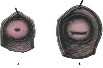
Fig. 329. Vaginal part of the cervix uteri, portio vaginalis cervicis uteri:
A - nulliparous woman; B - multiparous woman
The Peritoneum covers the intestinal surface of the body of the uterus, the upper part of the cervix, and the posterior vaginal fornix, and then reflects onto the rectum, forming a deep rectouterine pouch, excavatio rectouterina (Douglas pouch). Under normal conditions, intestinal loops may descend into the rectouterine pouch, whereas in pathological states, serous fluid, Blood, or pus may accumulate within it (Douglas abscess), which must be taken into account in clinical practice. Palpation and puncture of the rectouterine pouch are performed via the posterior fornix.
From the anterior abdominal wall, the peritoneum descends into the lesser pelvis, covers the apex and posterior surface of the urinary bladder, and then extends onto the uterus, forming the vesicouterine pouch, excavacio vesicouterina.
A contrast radiograph reveals the uterine cavity, which appears triangular in shape, with the angles of its base extending into the uterine tubes and its apex leading into the cervical canal and Vagina.
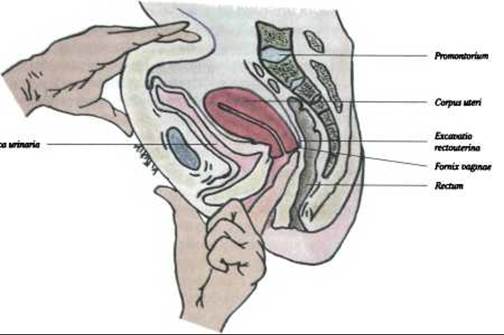
Fig. 330. Sagittal section through the female pelvis (diagram) (after H. Corning). A finger inserted into the vagina reaches the fornix vaginae
Last update: 08/08/2026
Editorial and Educational Adaptation: This material has been compiled based on the primary/original source text. The project team performed an editorial review, corrected technical inaccuracies, structured sections, and adapted the content for an educational format.
What was processed:
- elimination of formatting defects (OCR errors, structural breaks, corrupted characters);
- editorial organization of content;
- standardization of terminology in accordance with academic sources;
- verification of factual statements against the original source text.
All mentions of the author, publication year, and origin of the primary text have been preserved in accordance with the source.