Human Anatomy Part 1 - K. A. Dyubenko, A. K. Kolomiytsev, Yu. B. Chaikovsky 2002
Special Section
Urinary System, systema urinarium – General Overview
Urinary Organs – Urinary Bladder
The Urinary Bladder, vesica urinaria, is a hollow muscular organ that serves for the accumulation of urine and its periodic excretion through the Urethra (Fig. 312). The shape, size, and Location OF THE urinary bladder depend on the degree of its filling with urine and the condition of adjacent Organs. A full bladder is ovoid in shape, protrudes above the symphysis pubis, lies against the abdominal wall, and displaces the Peritoneum upward. This feature is taken into account in clinical practice when performing a puncture through the muscular layer of the abdominal wall to drain free fluid from the Abdominal cavity in cases of ascites, ostites. The capacity of the urinary bladder is 600-700 ml.
The following PARTS OF THE urinary bladder are distinguished:
- the apex, apex vesicae [vesicalis];
- the fundus, fundus vesicae;
- the body, corpus vesicae;
- the neck, cervix vesicae.
The apex of the urinary bladder faces upward and forward, while the fundus faces backward and downward. The body is located between the apex and the fundus, and the neck is situated at the transition of the bladder into the urethra. In addition, the following surfaces are distinguished: anterior, superior, posterior, and lateral. In children, the bladder is more elongated and has a spindle-shaped form. The urinary bladder is fixed in place by Ligaments and Muscles. Extending from the apex of the bladder to the umbilicus is the median umbilical ligament, lig. umbilicale medianum, which represents the obliterated embryonic duct, urachus. Furthermore, the urinary bladder is secured by smooth muscles: the pubovesical Muscle, m. pubovesicalis, attached to the Pubic Symphysis, and the rectovesical muscle, m. rectovesicalis, attached to the rectum.
Class="center">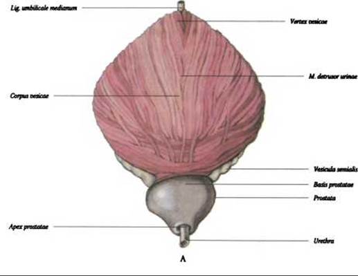
Fig. 312. Urinary bladder, vesica urinaria: A - anterior surface
Last update: 08/08/2026
Editorial and Educational Adaptation: This material has been compiled based on the primary/original source text. The project team performed an editorial review, corrected technical inaccuracies, structured sections, and adapted the content for an educational format.
What was processed:
- elimination of formatting defects (OCR errors, structural breaks, corrupted characters);
- editorial organization of content;
- standardization of terminology in accordance with academic sources;
- verification of factual statements against the original source text.
All mentions of the author, publication year, and origin of the primary text have been preserved in accordance with the source.