Human Anatomy Part 1 - K. A. Dyubenko, A. K. Kolomiytsev, Yu. B. Chaykovsky 2002
Special Part
Myology, myologia [the study of muscles] — Muscles and fascia of the upper extremity
Fasciae, synovial tendon sheaths, and fibrous canals
The following fascias are distinguished in the upper limb: the deltoid fascia, axillary fascia, brachial fascia, forearm fascia, and hand fascia.
1. Deltoid fascia, fascia deltoidea — formed by two layers (superficial and deep): the superficial layer covers the deltoid Muscle and passes along the anterior margin of the muscle into the pectoral fascia; the deep layer surrounds the deltoid muscle, separating it from the other Muscles of the shoulder girdle, and continues into the fascia covering the triceps muscle.
2. Axillary fascia, fascia axillaris — covers the axillary fossa from below, continues superiorly into the deltoid fascia, inferiorly into the brachial fascia, and posteriorly into the fascia covering the latissimus dorsi and teres major muscles.
3. Brachial fascia, fascia brachii [brachialis] — continues superiorly into the deltoid and axillary fascias, and forms fascial sheaths for the triceps brachii, biceps brachii, coracobrachialis, and brachialis muscles.
At the medial and lateral margins of the distal half of the arm, along the line separating the flexors from the extensors, the fascia sends off intermuscular septa: the medial intermuscular septum separates the medial HEAD of the triceps from the brachialis and pronator teres muscles; the lateral intermuscular septum separates the lateral and medial heads of the triceps from the brachialis and brachioradialis muscles.
4. Forearm fascia, fascia antebrachii — is a direct continuation of the brachial fascia. Throughout its extent, the fascia gives off septa that pass between individual muscle groups, forming sheaths for them. Proximally, the fascia fuses with the bicipital aponeurosis, while distally it forms bundles that encompass the radiocarpal joint region. In the upper half of the forearm, the fascial septa and interosseous membrane form three osteofascial muscle compartments:
1) the lateral compartment contains the brachioradialis and radial wrist extensor muscles;
2) the posterior compartment contains the extensor digitorum, extensor digiti minimi, ulnar wrist extensor, anconeus, and supinator muscles;
3) the anterior compartment, which is subdivided into a superficial layer containing the pronator teres, radial wrist flexor, flexor digitorum superficialis, palmaris longus, and ulnar wrist flexor muscles, and a deep layer containing the flexor digitorum profundus and flexor pollicis longus muscles.
In the wrist region, the forearm fascia thickens, forming the flexor and extensor retinacula on the PALMAR AND DORSAL surfaces, respectively: the flexor retinaculum (LNA) and extensor retinaculum for the flexor and extensor tendons. The flexor and extensor retinacula are crucial for stabilizing the positions of the tendons passing beneath them, especially during flexion and extension of the hand.
5. Hand fascias — are continuations of the forearm fascias. On the palmar surface of the hand, superficial and deep fascias are distinguished. The superficial fascia covers the muscles of the thenar and hypothenar eminences. In the central region, it thickens and continues into the palmar aponeurosis. The palmar aponeurosis, aponeurosis palmaris, is a thick, dense triangular tendinous plate whose base faces the fingers and whose apex is directed toward the wrist. The apex of the aponeurosis fuses with the palmar carpal ligament, the tendon of the palmaris longus muscle, and the flexor retinaculum. The distal part of the aponeurosis divides into four longitudinal bands directed toward the bases of digits II–V, where the longitudinal fibers weave into the anterior walls of the fibrous digital sheaths of the hand, vaginae fibrosae digitorum manus. The majority of the superficial bundles of the longitudinal aponeurotic fibers, distal to the transverse Skin creases of the palm, firmly fuse with the skin. A smaller portion extends deep between the digital flexor tendons and lumbrical muscles, participating in The formation of the sagittal septa of the palm (V. A. Klepikov, 1955).
The deep fascia covers the palmar interosseous muscles. On the palmar surface, the fascia forms five synovial sheaths (Fig. 165): the tendon Sheath of the long flexor of the thumb (Vagina tendinis m. flexoris pollicis longi), the common flexor sheath (vaginae communis musculorum flexorum), and the synovial sheaths of the tendons of digits II–IV (vaginae synovialis digitorum manus). It originates near the lower margin of the pronator quadratus muscle. The largest of these is the common sheath of the superficial and deep finger flexors. Its distal part continues into the synovial sheath of the fifth digit. In clinical practice, it is referred to as the ulnar bursa. Isolated synovial sheaths of the digits are located on the palmar surface of digits II–IV. In addition to those described, various anatomical variations of the flexor tendon synovial sheaths are encountered (Fig. 166).
Class="center">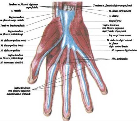
Fig. 165. Synovial sheaths of the hand — palmar surface (after V. P. Vorobyov)
The synovial sheath of the tendon of the long flexor of the thumb (radial bursa) is quite long. It originates at the level of the lower border of the pronator quadratus muscle, passes through the carpal canal (canalis carpi), and on the palmar surface lies between the superficial and deep heads of the short flexor of the thumb, reaching the distal phalanx. Within the carpal canal, the synovial sheaths (radial and ulnar) lie in close proximity to each other, and in 10% of cases communicate with one another, which, in the event of inflammation of either sheath, leads to The Development of a crossed or V-shaped Phlegmon (B. V. Ognev, 1960).

Fig. 166. Anatomical variations of the palmar tendon sheaths of the hand (after Lantz and Wachsmuth)
On the dorsal surface of the hand lies the dorsal Fascia of the hand (fascia dorsalis manus), which is divided into a superficial layer covering the extensor tendons and a deep layer covering the dorsal interosseous muscles. The extensor retinaculum (retinaculum extensorum) is a thickening of the fascia, about 2 cm wide, stretched between the distal ends of the radius and ulna. The extensor tendons of the hand and digits pass between the retinaculum (posteriorly) and the forearm bones and interosseous membrane (anteriorly). The extensor retinaculum splits into layers, forming fibrous canals for the tendons. The tendons passing through these canals are enclosed within synovial sheaths.
On the dorsal surface of the hand, the fascia forms six fibrous canals containing 11 tendons arranged in 8 synovial sheaths (Fig. 167):
- The first fibrous canal is located on the lateral surface of the radius. It contains two synovial sheaths: the tendon sheaths of the abductor pollicis longus and extensor pollicis brevis muscles (vaginae tendinum mm. abductoris longi et extensoris pollicis brevis) for the tendons of the abductor pollicis longus and extensor pollicis brevis.
- The second fibrous canal is located on the radius, distal to the muscular belly of the short flexor of the thumb. It transmits: laterally, the tendon of the extensor carpi radialis longus, and medially, the tendon of the extensor carpi radialis brevis, enclosed within the synovial sheaths of the radial wrist extensors (vaginae tendinum mm. extensorum carpi radialium).
- The third fibrous canal is located medial to the second and transmits the tendon of the extensor pollicis longus, which is enclosed in the synovial sheath of the extensor pollicis longus tendon (vagina tendinis m. extensoris pollicis longi).
- The fourth fibrous canal is located more medially than the previous one, along the medial margin of the radius. It transmits four tendons of the finger extensors and one tendon of the extensor indicis. All these tendons are enclosed within a single common synovial sheath (vagina tendinum mm. extensoris digitorum et extensoris indicis).
- The fifth fibrous canal is formed by a splitting of the Deep Layer of the extensor retinaculum and is located along the lateral margin of the distal radioulnar joint. It transmits the tendon of the extensor digiti minimi, enclosed within the synovial sheath of the extensor digiti minimi tendon (vagina tendinis m. extensoris digiti minimi).
- The sixth fibrous canal is located on the posteromedial surface of the ulna within its groove. It transmits the tendon of the extensor carpi ulnaris and its synovial sheath (vagina tendinis m. extensoris carpi ulnaris).

Fig. 167. Synovial sheaths of the hand — dorsal surface (after V. P. Vorobyov)
Last update: 08/08/2026
Editorial and Educational Adaptation: This material has been compiled based on the primary/original source text. The project team performed an editorial review, corrected technical inaccuracies, structured sections, and adapted the content for an educational format.
What was processed:
- elimination of formatting defects (OCR errors, structural breaks, corrupted characters);
- editorial organization of content;
- standardization of terminology in accordance with academic sources;
- verification of factual statements against the original source text.
All mentions of the author, publication year, and origin of the primary text have been preserved in accordance with the source.