Human Anatomy Part 1 - K. A. Dyubenko, A. K. Kolomiytsev, Yu. B. Chaykovsky 2002
Special Part
Myology, myologia [the study of muscles] - Muscles and fascia of the upper limb
Muscles of the upper limb, musculi membri superioris - Muscles of the pectoral girdle
The Muscles of the Upper Limb are divided into the muscles of the shoulder girdle and the free upper limb (arm, forearm, and hand).
1. Deltoid Muscle, m. deltoideus.
2. Supraspinatus muscle, m. supraspinatus.
3. Infraspinatus muscle, m. infraspinatus.
4. Teres minor muscle, m. teres minor.
5. Teres major muscle, m. teres major.
6. Subscapularis muscle, m. subscapularis.
1. Deltoid muscle, m. deltoideus — located subcutaneously in the deltoid region above the shoulder joint (Fig. 152). It is triangular in shape, with its apex pointing toward the humerus; its anterior border neighbors the m. pectoralis major and is separated from it by the sulcus deltoideopectoralis. The muscle originates from the lateral surface of the clavicle, the acromion of the scapula, and the spine of the scapula, correspondingly comprising three parts: pars clavicularis, pars acromialis, and pars spinalis. Its fascicles run laterally and inferiorly, inserting into the tuberositas deltoidea of the humerus.
Class="center">
Fig. 152. Muscles of the shoulder girdle and arm — posterior view (after V. P. Vorobyov)
Function: The deltoid muscle significantly contributes to the stabilization of the shoulder joint. The pars clavicularis flexes the arm anteriorly and medially rotates it slightly; the pars acromialis abducts the arm laterally up to the horizontal plane, while the pars spinalis extends the arm posteriorly and laterally rotates it slightly.
Blood supply: aa. circumflexa humeri posterior, thoraco acromialis, profunda brachii.
Innervation: n. axillaris (C V-C VI).
2. Supraspinatus muscle, m. supraspinatus, occupies the fossa of the same name on the scapula and is covered by the m. trapezius (Fig. 153). It originates from the walls of the fossa supraspinata and the supraspinous fascia, passes beneath the acromion and the lig. coracoacromiale, and inserts into the anterior part of the greater tubercle of the humerus and the capsule of the shoulder joint.
Function: Abducts the arm together with the deltoid muscle and tightens the capsule of the shoulder joint.
Blood supply: aa. suprascapularis, circumflexa scapulae.
Innervation: n. suprascapularis (C V-C VI).
3. Infraspinatus muscle, m. infraspinatus — covered by the deltoid and trapezius muscles (see Fig. 153). It originates from the fossa infraspinata and the infraspinous fascia, inserting into the middle part of the greater tubercle of the humerus and the capsule of the shoulder joint.
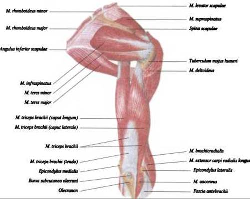
Fig. 153. Muscles of the shoulder girdle and arm — posterior view (after R. D. Sinelnikov)
Function: Extends the elevated arm posteriorly and laterally rotates the humerus; prevents pinching of the Joint Capsule.
Blood supply: aa. circumflexa scapulae, suprascapularis.
Innervation: n. suprascapularis (C V-C VI).
4. Teres minor muscle, m. teres minor — covered by the m. deltoideus (see Fig. 153). It originates on the dorsal aspect of the scapula from the lateral border and inserts into the posterior part of the greater tubercle of the humerus.
Function: Laterally rotates the arm and pulls back the capsule of the shoulder joint.
Blood supply: a. circumflexa scapulae.
Innervation: n. axillaris (C V–C VI).
5. Teres major muscle, m. teres major – located inferior to the teres minor and anterior to the triceps brachii muscle (see Fig. 153). In a living person, this muscle is easily palpable beneath the Skin when the arm is abducted and pressed against the trunk. It originates from the lower part of the lateral border of the scapula and the infraspinous fascia, and inserts into the crista tuberculi minoris.
Function: medial rotation and adduction of the arm.
Blood supply: a. subscapularis.
Innervation: n. subscapularis (C V–C VII).
6. Subscapularis muscle, m. subscapularis – situated on the anterior surface of the scapula (Fig. 154), it is a multipennate muscle with considerable lifting power.

Fig. 154. Subscapularis muscle, n. subscapularis, and teres major muscle, n. teres major – anterior view (after Spalteholz)
It originates from the subscapular fossa and the corresponding fascia, and inserts into the lesser tubercle of the humerus and the capsule of the shoulder joint.
Function: medially rotates and adducts the arm; acting independently, it acts as a pronator of the arm.
Blood supply: a. subscapularis.
Innervation: n. subscapularis.
Last update: 08/08/2026
Editorial and Educational Adaptation: This material has been compiled based on the primary/original source text. The project team performed an editorial review, corrected technical inaccuracies, structured sections, and adapted the content for an educational format.
What was processed:
- elimination of formatting defects (OCR errors, structural breaks, corrupted characters);
- editorial organization of content;
- standardization of terminology in accordance with academic sources;
- verification of factual statements against the original source text.
All mentions of the author, publication year, and origin of the primary text have been preserved in accordance with the source.