Human Anatomy, Part 1 - K. A. Diubenko, A. K. Kolomiitsev, Yu. B. Chaikovsky 2002
Special Section
Myology, myologia [the study of muscles] — Muscles and Fasciae of the Neck
Muscles of the neck, musculi colli [cervicis] — Middle muscles of the neck — Suprahyoid muscles, mm. suprahyoidei
1. Digastric Muscle, m. digastricus, lies beneath the Mandible (Fig. 142 A). The posterior belly, venter posterior, originates from the mastoid notch of the Temporal bone and is covered by the m. sternocleidomastoideus. The anterior belly, venter anterior, originates from the digastric fossa of the mandible. Both bellies converge and attach by a tendon to the body of the Hyoid bone (os hyoideum).
Function: with the hyoid bone fixed, it depresses the mandible; with the mandible fixed, it draws the hyoid bone upward.
Blood supply: anterior belly – a. submentalis; posterior belly – aa. occipitalis, auricularis posterior.
Innervation: anterior belly – n. mylohyoideus (n. trigeminus (V)); posterior belly – r. digastricus (n. facialis (VII)).
2. Stylohyoid muscle, m. stylohyoideus, is spindle-shaped and located superior to the posterior belly of the m. digastricus (see Fig. 142A). It originates from the lateral aspect of the Base of the styloid process (processus styloideus), passes downward and forward toward the hyoid bone, and inserts at the junction of the body and the greater horn of the hyoid bone.
Function: displaces the hyoid bone backward and upward.
Blood supply: aa. occipitalis, facialis, r. suprahyoideus, a. lingualis.
Innervation: r. stylohyoideus (n. facialis (VII)).
3. Geniohyoid muscle, m. geniohyoideus, is triangular in shape and situated superior to the preceding muscle (see Fig. 142B). It originates from the mental spine (spina mentalis), extends posteriorly, and inserts into the anterior surface of the body of the hyoid bone (corpus ossis hyoidei).
Function: draws the hyoid bone upward and forward; with the hyoid bone fixed, it assists in depressing the mandible.
Blood supply: aa. sublingualis, submentalis.
Innervation: ansa cervicalis.
4. Mylohyoid muscle, m. mylohyoideus, appears as a thin sheet (see Fig. 142A) located between the mandible and the hyoid bone. Together with its counterpart from the opposite side, it closes the space between the mandible and the hyoid bone, forming the oral Diaphragm, which serves as the floor of the Oral Cavity. The muscle fibers originate from the mylohyoid line (linea mylohyoidea). Along the midline, where the fibers from both sides meet, runs a fibrous raphe. The posterior fibers attach to the anterior surface of the body of the hyoid bone.
Class="center">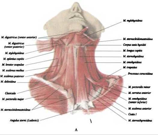
Fig. 142. Muscles of the neck: suprahyoid and Infrahyoid muscles — anterior view (after V. P. Vorobyov)
Function: with the mandible fixed, it elevates and pulls the hyoid bone upward and forward; with the hyoid bone fixed, it assists in depressing the mandible.
Blood supply: aa. sublingualis, submentalis.
Last update: 08/08/2026
Editorial and Educational Adaptation: This material has been compiled based on the primary/original source text. The project team performed an editorial review, corrected technical inaccuracies, structured sections, and adapted the content for an educational format.
What was processed:
- elimination of formatting defects (OCR errors, structural breaks, corrupted characters);
- editorial organization of content;
- standardization of terminology in accordance with academic sources;
- verification of factual statements against the original source text.
All mentions of the author, publication year, and origin of the primary text have been preserved in accordance with the source.