MEDICAL BIOLOGY, HUMAN ANATOMY, PHYSIOLOGY AND PATHOLOGY - Y.I. Fedonyuk 2010
ANATOMY, PHYSIOLOGY, PATHOLOGY
CHAPTER 7. THE NUTRITIONAL PROCESS AND ITS PATHOLOGY
PATHOLOGY OF THE DIGESTIVE SYSTEM
LIVER PATHOLOGY
2. HEPATOSIS
Hepatosis is a Liver disease characterized by dystrophy and necrosis of hepatocytes; it can be either hereditary or acquired, and may have an acute (Toxic Liver Dystrophy) or chronic (fatty liver) course.
Hereditary hepatoses develop due to impaired METABOLISM of fats (lipidoses), CARBOHYDRATES (glycogenoses), pigments, and minerals. Many hereditary hepatoses are storage diseases that ultimately result in liver cirrhosis.
Toxic liver dystrophy is an acute (rarely chronic) condition driven by progressive massive necrosis of liver Cells, leading to rapidly developing acute Liver failure. Massive liver necrosis most commonly occurs under The Influence of exogenous intoxications (poisoning by mushrooms, spoiled food, chemicals, medications) and endogenous intoxications (Pregnancy toxemia, thyrotoxicosis).
The pathomorphological Changes in the liver vary across different Stages of the disease, which typically lasts about three weeks. Early in the disease, the liver enlarges and takes on a bright yellow color due to fatty degeneration of hepatocytes; by the end of the second week, its size decreases. Microscopically, fatty degeneration at this stage gives way to necrosis and autolysis, resulting in The formation of lipid-protein detritus (the stage of yellow liver atrophy). Subsequently, the stroma collapses and sinusoids become engorged with Blood (the stage of red atrophy). At the peak of the illness, jaundice and hemorrhages in the Skin and mucous membranes appear. Patients typically die from hepatic or hepatorenal failure. The ultimate consequence of toxic dystrophy is postnecrotic liver cirrhosis.
Fatty hepatosis (Fatty liver disease) is a chronic liver condition characterized by the excessive accumulation of fat within liver cells.
Chronic alcohol intoxication plays a primary role in The Development of fatty hepatosis.
In fatty hepatosis, the liver is enlarged, yellow or red-brown in color, and has a smooth surface. Fat accumulation is visible within hepatocytes. In cases of severe fatty infiltration, liver cells perish, and fat droplets coalesce to form intracellular fat cysts; a cellular reaction develops around these cysts, accompanied by Connective Tissue proliferation (Fig. 7.32).
Class="center">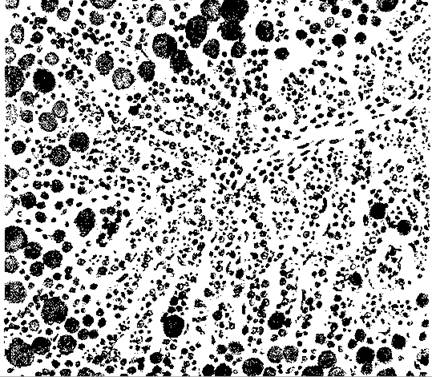
Fig. 7.32. Fatty degeneration of the liver
Last update: 08/08/2026
Editorial and Educational Adaptation: This material has been compiled based on the primary/original source text. The project team performed an editorial review, corrected technical inaccuracies, structured sections, and adapted the content for an educational format.
What was processed:
- elimination of formatting defects (OCR errors, structural breaks, corrupted characters);
- editorial organization of content;
- standardization of terminology in accordance with academic sources;
- verification of factual statements against the original source text.
All mentions of the author, publication year, and origin of the primary text have been preserved in accordance with the source.