MEDICAL BIOLOGY, HUMAN ANATOMY, PHYSIOLOGY AND PATHOLOGY - Ya.I. Fedoniuk 2010
ANATOMY, PHYSIOLOGY, PATHOLOGY
CHAPTER 7. THE NUTRITIONAL PROCESS AND ITS PATHOLOGY
ANATOMY AND PHYSIOLOGY OF THE DIGESTIVE ORGANS
PHARYNX
The Pharynx is a part of the alimentary canal connecting the Oral Cavity to the Esophagus, as well as a section of the respiratory tract connecting the Nasal cavity to the Larynx. The pharyngeal cavity is the site where the digestive and respiratory pathways cross (Fig. 7.7).
Class="center">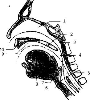
Fig. 7.7. Diagram of the respiratory (indicated by a solid line) and digestive (indicated by a dashed line) pathways in the pharyngeal region and their intersection
The pharynx lies posterior to the nasal cavity, oral cavity, and larynx, and anterior to the basilar part of the Occipital bone and the first six cervical vertebrae. Accordingly, it is divided into three parts: the nasal part (nasopharynx), the oral part (oropharynx), and the laryngeal part (laryngopharynx).
The pharyngeal cavity (Fig. 7.8) communicates with:
1) the nasal cavity through the choanae;
2) the tympanic cavity (Middle ear) through the openings of the auditory (Eustachian) tubes located on the lateral wall of the pharynx;
3) the oral cavity through the fauces;
4) the larynx through the laryngeal inlet;
5) the esophagus (the pharynx directly continues into the esophagus).
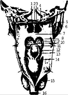
Fig. 7.8. Pharyngeal cavity; posterior view
In the region of the pharynx, there are accumulations of lymphoid tissue that form the Tonsils. They are situated at the entrance to the pharynx and form an almost complete ring known as the Pirogov-Waldeyer ring. These include: the pharyngeal tonsil (1), the tubal tonsils (2), the palatine tonsils (2), and the lingual tonsil (1).
The framework of the pharyngeal wall is formed by a fibrous plate attached superiorly to the external Base of the Skull. Externally, this plate is covered by Muscles, and internally by the mucous membrane. The pharyngeal muscles are divided into longitudinal and circular layers (Fig. 7.9). The longitudinal muscles include the stylopharyngeus and palatopharyngeus, which elevate the pharynx. The circular muscles comprise three constrictors: the superior, middle, and inferior pharyngeal constrictors. Their contraction propels the food bolus down into the esophagus.

Fig. 7.9. Muscles of the pharynx; posterior view
Externally, the muscular layer is covered by loose Connective Tissue—the adventitia.
Last update: 08/08/2026
Editorial and Educational Adaptation: This material has been compiled based on the primary/original source text. The project team performed an editorial review, corrected technical inaccuracies, structured sections, and adapted the content for an educational format.
What was processed:
- elimination of formatting defects (OCR errors, structural breaks, corrupted characters);
- editorial organization of content;
- standardization of terminology in accordance with academic sources;
- verification of factual statements against the original source text.
All mentions of the author, publication year, and origin of the primary text have been preserved in accordance with the source.