HUMAN MEDICAL BIOLOGY, ANATOMY, PHYSIOLOGY, AND PATHOLOGY - Y.I. Fedoniuk 2010
ANATOMY, PHYSIOLOGY, PATHOLOGY
CHAPTER 5. BLOOD AND LYMPH CIRCULATION PROCESSES AND THEIR PATHOLOGY
CARDIOVASCULAR SYSTEM DISEASES
HEART DEFECTS
The term "Heart defect" has long been used to denote congenital or acquired morphological Changes in the valvular apparatus, cardiac septa, or the major vessels extending from The Heart.
Congenital heart defects encompass Disorders of the heart and great vessels characterized by abnormal positioning and morphological Structure resulting from impaired or incomplete development during the intrauterine, or less commonly, postnatal developmental periods. This pathology is one of the most common and severe cardiovascular diseases.
Acquired heart defects are conditions that develop during a patient's lifetime As a result of diseases or traumatic injuries to the heart. The causes of acquired heart defects are highly diverse, though the most frequent is rheumatic fever (accounting for at least 90 % of all cases) (Fig. 5.55).
Class="center">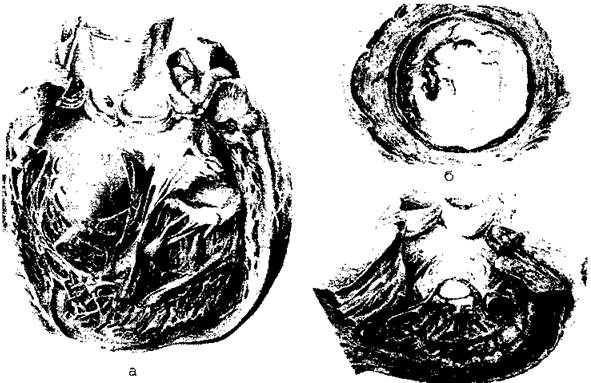
Fig. 5.55. Rheumatic heart defects
In certain heart defects, an obstacle in the form of a narrowing (bottleneck) impedes Blood outflow—for example, stenosis of the mitral orifice or aortic orifice, or narrowing of the Thoracic Aorta in coarctation. In such cases, blood must pass through a severely restricted opening. As a result, during physical exertion, a patient with stenosis shows virtually no increase in Cardiac Output because the heart is unable to overcome the sharply increased resistance.
In valvular insufficiency, the mechanisms of hemodynamic disturbance are different. Due to the incomplete closure of the valve cusps, a portion of the blood flows backward during systole: in mitral or tricuspid insufficiency, from the ventricles into the atria; and in aortic or pulmonary valve insufficiency, from the vessels back into the corresponding ventricle. The volume of this regurgitant blood flow is determined by the pressure gradient between the chambers and the size of the residual gap remaining after the cusps close.
Thus, in stenosis, the heart's excess work is expended on overcoming resistance, which is achieved by increasing pressure. In valvular insufficiency, the increased Energy Expenditure of the heart is caused by pumping an additional volume of blood. Consequently, stenosis results in systolic overload, or isometric hyperfunction, whereas valvular insufficiency results in diastolic overload, or isotonic hyperfunction.
Hemodynamic disturbances are particularly unfavorable when stenosis and valvular insufficiency are combined. In this scenario, a portion of the blood is ejected backward into the atrium during systole, and subsequently, this same portion of blood must pass through the narrowed orifice all over again. This creates conditions far more complex than those seen in an isolated defect, whether stenosis or insufficiency.
Hemodynamic alterations in many congenital defects are caused by abnormal communications between the SYSTEMIC AND PULMONARY circulations, as well as between cardiac chambers (via septal defects) or major vessels (via a patent ductus arteriosus or aortopulmonary fistula). If There is a pressure differential between connected sections of The Cardiovascular system, a blood shunt occurs, meaning blood flows from one chamber into another.
Arterial blood is located in the left heart chambers, while venous blood is found in the right chambers. The mixing of arterial blood into the venous blood is termed a left-to-right shunt, whereas the mixing of venous blood into the arterial blood is termed a right-to-left shunt.
When a shunt is present, the cardiac output circulating through the systemic and pulmonary circulations will differ. In a left-to-right shunt, the cardiac output of the systemic Circulation is lower than that of the Pulmonary Circulation, while in a right-to-left shunt, it is higher.
Blood shunts affect the workload of the heart, which under these conditions performs excess (unproductive) labor.
This additional work leads to organ hypertrophy. In heart defects, hypertrophy is observed in those chambers subjected to increased load, alongside some degree of atrophy in the sections performing less work.
Within certain limits, myocardial hypertrophy is a beneficial process, but excessive development leads to sclerotic changes and diminishes contractile capacity. Furthermore, when myocardial thickening becomes overly pronounced, the amplitude of its contractions drops, and the cardiac wall becomes rigid. Its ability to expand during diastole and reduce cavity volume during systole declines. These phenomena explain the clinical course of heart defects, wherein hemodynamic disturbances are initially compensated before eventually progressing to decompensation with The Development of circulatory failure.
Last update: 08/08/2026
Editorial and Educational Adaptation: This material has been compiled based on the primary/original source text. The project team performed an editorial review, corrected technical inaccuracies, structured sections, and adapted the content for an educational format.
What was processed:
- elimination of formatting defects (OCR errors, structural breaks, corrupted characters);
- editorial organization of content;
- standardization of terminology in accordance with academic sources;
- verification of factual statements against the original source text.
All mentions of the author, publication year, and origin of the primary text have been preserved in accordance with the source.