HUMAN MEDICAL BIOLOGY, ANATOMY, PHYSIOLOGY, AND PATHOLOGY - Ya.I. Fedonyuk 2010
BIOLOGY
CHAPTER 1. BIOLOGICAL FOUNDATIONS OF HUMAN VITAL ACTIVITY
1.4. ONTOGENETIC LEVEL OF LIFE ORGANIZATION
1.4.2. Basics of Human Genetics
Genealogical Method
The term "genealogy" (genealogia) comes from Greek and means "pedigree". The genealogical method is a method of studying human heredity through family trees. It was proposed by the English scientist F. Galton (1865). The Essence of the method consists in establishing kinship relations and tracing a studied trait (disease) among close and distant, direct and collateral relatives. The genealogical method is the most universal method in Human Genetics. Technically, it consists of two stages: 1) constructing a pedigree and 2) genealogical Analysis of the pedigree.
Constructing a Pedigree.
The Construction of a pedigree begins with the proband. A proband is the individual whose pedigree needs to be compiled and analyzed. Most often, this is a patient or a carrier of the Gene for the studied trait. Children of the same parental pair are called sibs (brothers and sisters). Usually, a pedigree is compiled for one or a few traits. It is technically impossible to compile it for all known traits. When constructing a pedigree, symbols proposed in 1931 by G. Just are used (Fig. 1.68).
Class="center">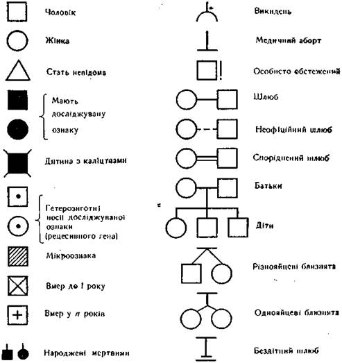
Fig. 1.68. Genetic symbols for constructing a pedigree chart
Rules for constructing pedigrees:
1) the proband on the pedigree chart is indicated by an arrow; 2) individuals of the same generation occupy a separate row or circle; 3) generations are designated on the left by Roman numerals; the oldest generation is placed at the top of the pedigree and designated by the numeral I, while the youngest
- at the bottom of the pedigree; 4) all members of the same generation are placed in order of birth (from left to right) horizontally and designated by Arabic numerals.
To ensure the reliability of the analysis results, at least 3-4 generations should be covered. The pedigree includes all family members: healthy, sick, stillborn and miscarried, defective, and mentally disabled. The more reliable information available about the health of the proband's relatives, the more informative the genealogical analysis will be. When referring to any family member, the generation number is indicated first, followed by the family member in that same generation: II-3 or III-7. A systematic description of information about the proband and their relatives (the legend) must be attached to the pedigree chart. The legend must reflect the following data: 1) results of clinical and paraclinical examination of the proband; 2) information from personal examination of the proband's relatives; 3) comparison of the results of the proband's personal examination with data from interviewing their relatives; 4) written information about relatives living in another locality.
Genealogical Analysis of a Pedigree. Pedigree analysis allows one to establish: 1) The Nature of the trait (hereditary or non-hereditary); 2) the type of inheritance; 3) the zygosity of the proband (homozygous or heterozygous) for the studied trait; 4) the probability risk of the hereditary trait manifesting in descendants; 5) given a pedigree including the maximum number of relatives, it is also possible to determine gene penetrance, analyze gene linkage, perform chromosome mapping, and study the intensity of the Mutational Process and mechanisms of gene interaction.
The first task of genealogical analysis is to establish whether the studied trait is hereditary or non-hereditary. If the studied trait (or disease) occurs several times in a pedigree, its hereditary nature can be suspected. However, the possibility of phenocopies must be excluded. For example, if the same harmful factor acted on a woman during all her pregnancies, she may give birth to children with identical anomalies. Another example: the same occupational hazards can cause similar diseases in members of the same family. After establishing the hereditary Nature of the trait (disease), it is necessary to determine the type of inheritance: autosomal dominant, autosomal recessive, X-linked dominant, X-linked recessive, and Y-linked.
The autosomal dominant type of inheritance is caused by the transmission over a series of generations of a dominant gene localized in an autosome. This type of inheritance is characterized by: 1) with a sufficient number of offspring, the trait (disease) manifests in every generation (vertical inheritance in the pedigree); 2) males and females inherit the trait with equal frequency; 3) both parents transmit the trait to their children to an equal degree; 4) one or both parents of a sick child are sick; 5) the probability of giving birth to a sick child in a family where one parent is sick and heterozygous while the other is healthy (Aa x aa) is 50% (Tab. 1.8).
Table 1.8.
Autosomal dominant type of inheritance
Parents' genotypes |
Children's genotypes |
Healthy children, % |
Sick children, % |
АА х АА |
АА |
- |
100 |
АА х Аа |
АА, Аа |
- |
100 |
Аа х Аа |
АА, Аа, Аа, аа |
25 |
75 |
Аа х аа |
Аа, аа |
50 |
50 |
аа х аа |
аа |
100 |
- |
The autosomal dominant type of inheritance can be illustrated by the example of brachydactyly (Fig. 1.69). If the brachydactyly gene is designated as A and the normal gene as a, we can conclude that all pedigree members with shortened fingers are heterozygous and have the Aa genotype. Since all individuals with brachydactyly in the analyzed pedigree married normal women or men, these marriages serve as an example of Mendelian test crossing Aa x aa. When counting all individuals with brachydactyly and with normal finger length from such marriages, it is evident that phenotypic segregation is close to 1:1. This pedigree confirms the applicability of G. Mendel's Laws to humans.
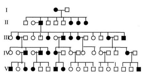
Fig. 1.69. Pedigree with Autosomal dominant inheritance (brachydactyly)
It must be taken into account that in autosomal dominant inheritance, a trait may be absent in one generation. This occurs when diseases do not manifest immediately, but at a certain
age, such as Huntington's disease (average onset at 38–40 years). In the case of death at an earlier age, there will be no data regarding a possible illness in that family member, but the probability of the disease appearing in descendants remains. There are mild or incomplete forms of disorders with low expressivity of the mutant gene, or the latter may be suppressed by some epistatic gene.
Autosomal Recessive Inheritance is caused by the transmission across generations of a recessive gene localized in an autosome. Typical features of this type include: 1) even with a sufficient number of offspring, the trait (disease) does not manifest in every generation; 2) siblings (brothers and sisters) are primarily affected (horizontal inheritance) rather than parents and children, as in autosomal dominant inheritance; 3) males and females inherit the trait with equal frequency; 4) the probability of having affected children increases in consanguineous marriages; 5) only recessive homozygotes (aa) are affected, whereas heterozygotes (Aa) are healthy but act as carriers of the pathological gene; 6) the probability of having affected children from healthy heterozygous parents (Aa x Aa) is 25% (Table 1.9).
Table 1.9.
Autosomal Recessive Inheritance
Parent Genotypes |
Offspring Genotypes |
Healthy Children, % |
Affected Children, % |
АА х АА |
АА |
100 |
- |
АА х Аа |
АА, Аа |
100 |
- |
Аа х Аа |
АА, Аа, Аа, аа |
75 |
25 |
Аа х аа |
Аа, аа |
50 |
50 |
аа х аа |
аа |
- |
100 |
An example of a pedigree with autosomal recessive inheritance is shown in Fig. 1.70. Pedigree analysis indicates that phenylketonuria is inherited as an autosomal recessive trait. Healthy parents in the third generation, who were first cousins, had affected daughters. Consequently, both parents were carriers of the disease (Aa), and the gene is recessive (a).

Fig. 1.70. Pedigree of a family with an autosomal recessive mode of inheritance (phenylketonuria)
X-linked dominant inheritance is caused by the localization of a dominant gene in the segment of the X chromosome that lacks a homolog on the Y chromosome. Key characteristics of this type include: 1) an affected father (XAY) will have all daughters affected, as they receive the paternal X chromosome carrying the dominant gene, while all sons will be born healthy because they inherit the maternal X chromosome; 2) if the mother is homozygous for the abnormal gene (XAXA), she will transmit the trait (disease) to all daughters and all sons; 3) if the mother is heterozygous, she will transmit the trait (disease) to half of her daughters (XAXa) and half of her sons (XAY); the probability of having healthy children is 50% regardless of sex.
Conditions inherited in an X-linked dominant manner include vitamin D-resistant Rickets and brown tooth enamel (Fig. 1.71).
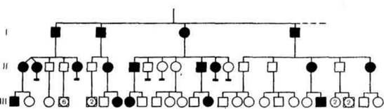
Fig. 1.71. Inheritance of an X-linked dominant trait (vitamin D-resistant rickets)
X-linked recessive inheritance. The recessive gene for the trait is localized in the segment of the X chromosome that has no homolog on the Y chromosome. Typical features of this inheritance pattern: 1) males are predominantly affected; 2) there is no transmission of the trait (disease) from father to son; 3) a female carrier (XAXa) transmits the disease to half of her sons, all daughters are healthy, but half of them are carriers;
4) all daughters of an affected father (XaY) will be phenotypically healthy, but heterozygous carriers;
5) if the mother is homozygous (XaXa) for this gene, all sons will be affected.
X-linked recessive disorders include hemophilia (p. 111), color blindness, Duchenne muscular dystrophy, and Lesch-Nyhan syndrome. Pedigrees with an X-linked recessive inheritance pattern show a predominance of affected males (Fig. 1.72).
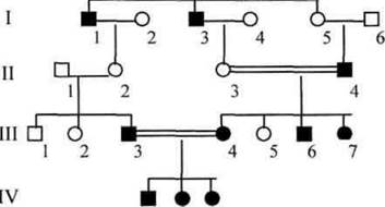
Fig. 1.72. X-linked recessive inheritance (color blindness).
Y-linked inheritance (holandric inheritance) is caused by the localization of a gene in the segment of the Y chromosome that has no homolog on the X chromosome. With this type, the trait is transmitted exclusively through the male line from generation to generation—from father to all his sons, since a father passes his Y chromosome only to his sons.
Y-linked traits include hypertrichosis of the ear rim ("hairy ears") and ichthyosis (excessive keratinization of the Skin—"fish scale disease"). An example of a pedigree is presented in Fig. 1.73.
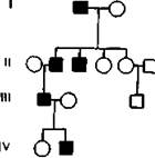
Fig. 1.73. Hypothetical pedigree for the inheritance of a Y-linked trait
Mitochondrial Inheritance. This is associated with genes located in Mitochondrial DNA. Cell/35.html">Mitochondria are inherited exclusively through the maternal line via the Cytoplasm of the egg Cells. Spermatozoa contain almost no cytoplasm. Various Mutations in Mitochondrial Genes have been described. Gene Mutations in mitochondrial DNA are detected in mitochondrial disorders—Leber's hereditary optic neuropathy, mitochondrial myopathies, progressive ophthalmoplegias, cardiomyopathies, and blindness-ataxia syndrome. The mitochondrial type of inheritance is characterized by the following features: 1) the disease is transmitted exclusively by the mother; both girls and boys can be affected; 3) affected fathers do not transmit the disorder to either daughters or sons (Fig. 1.74).
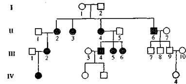
Fig. 1.74. Pedigree illustrating the transmission of a disorder via mitochondria.
Last update: 08/08/2026
Editorial and Educational Adaptation: This material has been compiled based on the primary/original source text. The project team performed an editorial review, corrected technical inaccuracies, structured sections, and adapted the content for an educational format.
What was processed:
- elimination of formatting defects (OCR errors, structural breaks, corrupted characters);
- editorial organization of content;
- standardization of terminology in accordance with academic sources;
- verification of factual statements against the original source text.
All mentions of the author, publication year, and origin of the primary text have been preserved in accordance with the source.