BIOLOGY Volume 2 - A Guide to General Biology - 2004
13. PLANT TRANSPORT
13.5. Water Uptake by Roots
Fig. 13.17 illustrates the Introduction/19.html">Primary Structure of a typical dicotyledonous ROOT.
Class="center">
Fig. 13.17. A. Anatomy of a young root of a typical dicotyledonous plant, buttercup (Ranunculus sp.). B. Light micrograph of a transverse section of a buttercup root under low magnification.
The bulk of Water is absorbed by the younger Regions of the root within the root Hair zone. As the root grows through the soil, new root hairs are continuously formed while older ones die off. These new root hairs develop a short distance behind the elongation zone. They appear as tubular outgrowths of epidermal Cells (Fig. 13.17) that dramatically increase the surface area available for the absorption of water and mineral salts, establishing intimate contact with soil particles.
Fig. 13.18, A schematically illustrates the pathways of water movement across the root. A water potential gradient exists within the root, running from a higher potential in the root Hair cells to a lower potential in the cells adjacent to the xylem. This gradient is maintained in two ways:
1) by the upward Movement of water through the xylem, which, as discussed earlier, generates tension (negative pressure) within the xylem and thereby lowers The water potential of the xylem sap;
2) because the osmotic potential of the xylem sap is lower (more negative) than that of the dilute soil solution.
Water moves across the root via the same pathways as in leaves, namely the apoplast, symplast, and vacuolar pathways.
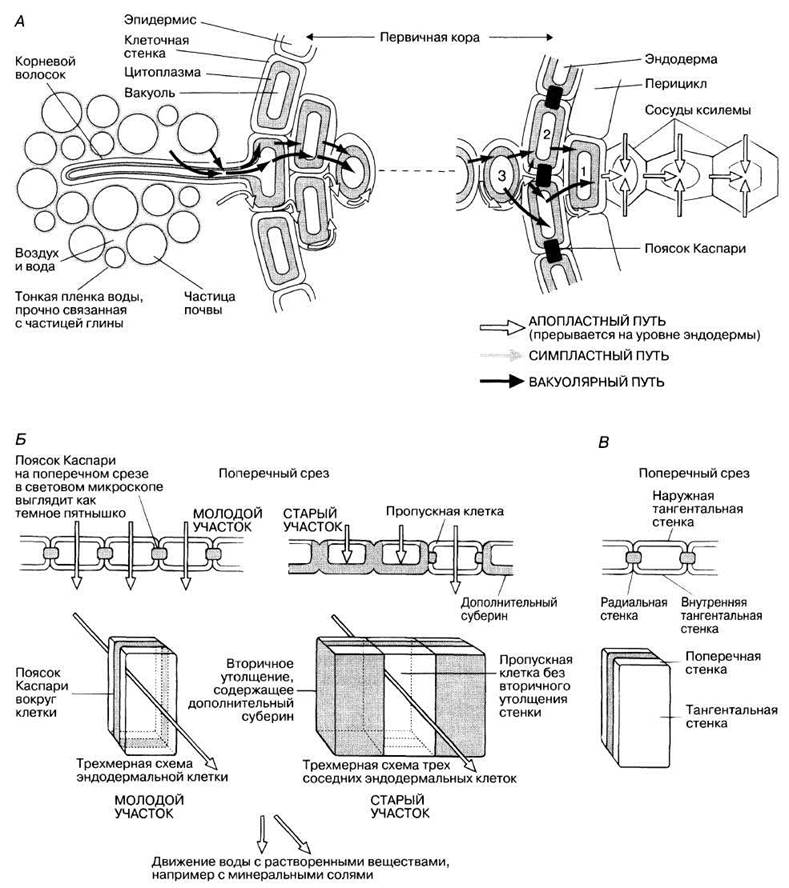
Fig. 13.18. A. Diagram of water and ion movement in a root (shown in transverse section). Cell wall thickness has been exaggerated for clarity. Cells 1, 2, and 3 are referenced in the text. For both water and dissolved solutes, the apoplastic pathway is predominant; the symplastic pathway plays a minor role, becoming essential only at the endodermis, while movement via the vacuolar pathway is negligible. B. Structure and function of the root endodermis. Young cells are characterized by Casparian strips, whereas older cells (except for passage cells) exhibit additional suberin deposition in their walls. C. Cell wall orientations: transverse and radial walls are anticlinal (i.e., perpendicular to the root surface), whereas the tangential wall is periclinal (i.e., parallel to the root surface).
13.5.1. Symplastic and Vacuolar Pathways
As water is drawn up the root xylem, it is replaced by water from surrounding parenchymal cells, such as cell 1 in Fig. 13.18, A. Consequently, the WATER POTENTIAL OF this cell drops, drawing water in from neighboring cell 2 via osmosis or directly through the symplast, as described in Section 13.3.2 for leaf mesophyll. This, in turn, lowers the water potential of cell 2, causing water to flow into it from cell 3, and so on across the entire root all the way to the epidermal root hairs.
The water potential of the soil solution is higher than that of the epidermal cells and root hairs. Consequently, water enters the root from the outside by osmosis.
13.17. Arrange the following components in order of decreasing water potential (using the > symbol): soil solution, xylem sap, cells 1, 2, and 3, root hair cell.
Last update: 06/08/2026
Editorial and Educational Adaptation: This material has been compiled based on the primary/original source text. The project team performed an editorial review, corrected technical inaccuracies, structured sections, and adapted the content for an educational format.
What was processed:
- elimination of formatting defects (OCR errors, structural breaks, corrupted characters);
- editorial organization of content;
- standardization of terminology in accordance with academic sources;
- verification of factual statements against the original source text.
All mentions of the author, publication year, and origin of the primary text have been preserved in accordance with the source.