Harper's Illustrated Biochemistry, Volume 1 - Murray R. 1993
Structure and Functions of Proteins and Enzymes
Proteins: Structure and Properties
Methods for Determining Primary Structure
First, Conjugated Proteins are stripped of their prosthetic groups (such as heme) and their Disulfide Bonds are oxidized, yielding linear Polypeptides (see Fig. 4.10). The sequencing Methods for these polypeptides were already discussed in Chapter 4. Most proteins contain only the Amino Acids listed in Table 3.3, though some proteins feature derivatives of these amino acids (Tables 5.4 and 5.5). Methods for identifying Amino Acid Derivatives fall outside The Scope of this chapter; we will simply note that their presence complicates Primary Structure determination.
Class="center">Table 5.4. Modification of a-COOH and a-NH2 groups within proteins 1)
|
а-СООН |
a-NH2 |
||
|
Amide |
N-formyl |
N-acetyl |
N-methyl |
|
Ala |
Ala |
||
|
Asp Glu |
Asp |
Asp |
|
|
Gly His |
Gly |
Gly |
Gly |
|
Met |
Met |
Met |
Met |
|
Phe |
|||
|
Pro |
Ser |
Ser |
|
|
Thr |
Thr |
||
|
Tyr Val |
Val |
1) Adapted, with kind permission From the Authors, from Uy R., Wold F.: Posttranslational Covalent Modification of proteins. Science: 1977: 198: 890.
Introduction/19.html">Primary structure of Insulin and Ribonuclease
Insulin consists of two polypeptide chains covalently linked by disulfide bonds (Fig. 5.9). The A-chain has a Gly residue at the N-terminus and an Asn residue at the C-terminus; in the B-chain, the N- and C-terminal residues are Phe and Ala, respectively. Oxidation of insulin with performic acid cleaves the disulfide bonds connecting the A- and B-chains. During Biosynthesis, both chains are initially part of a single proinsulin polypeptide chain, which undergoes post-synthesis proteolytic Processing to yield insulin (Chapter 51).
Table 5.5. Modified functional groups in amino acid side chains within proteins1)
|
—OH |
—N (in side chain) |
||
|
PO3H2 |
N-methyl |
N-dimethyl |
N-trimethyl |
|
Arg His |
Arg |
||
|
Ser |
Lys |
Lys |
Lys |
|
Thr |
|||
|
Tyr |
|||
1) Adapted, with kind permission from the authors, from Uy R., Wold F.: Posttranslational covalent modification of proteins. Science: 1977: 198: 890.
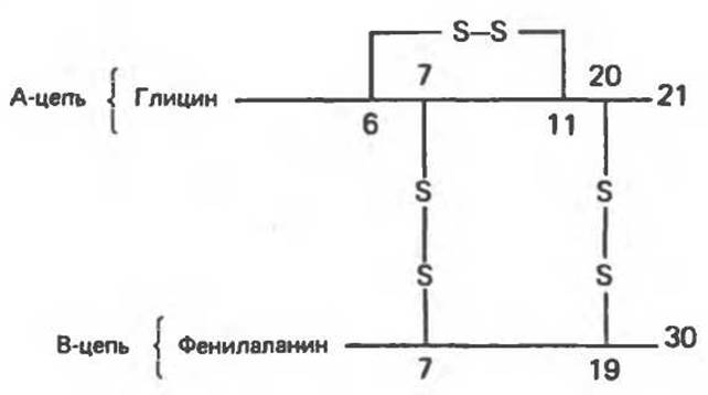
Fig. 5.9. Human insulin A- and B-chains connected by disulfide bonds.
Ribonuclease consists of a single chain of 124 residues, with Lys at the N-terminus and Val at the C-terminus. Eight Cysteine residues are joined by disulfide bonds, forming four cross-links within the protein (Fig. 5.10).
Last update: 06/08/2026
Editorial and Educational Adaptation: This material has been compiled based on the primary/original source text. The project team performed an editorial review, corrected technical inaccuracies, structured sections, and adapted the content for an educational format.
What was processed:
- elimination of formatting defects (OCR errors, structural breaks, corrupted characters);
- editorial organization of content;
- standardization of terminology in accordance with academic sources;
- verification of factual statements against the original source text.
All mentions of the author, publication year, and origin of the primary text have been preserved in accordance with the source.