Principles of Biochemistry, Volume 3 - A. Lehninger 1985
Molecular Mechanisms of Genetic Information Transfer
Protein Synthesis and Its Regulation
tRNA is required for amino acid activation
To understand how tRNAs act as adaptors in translating from the nucleic acid language to the protein language, we must first examine their Structure. tRNAs are relatively small, single-stranded molecules. Bacterial and extramitochondrial eukaryotic tRNAs consist of 73-93 NUCLEOTIDES, corresponding to a Molecular Weight of 24,000-31,000. (Cell/35.html">Mitochondria contain specialized tRNAs of a slightly smaller size.) Each amino acid corresponds to at least one tRNA; Some Amino Acids correspond to two or more specific tRNAs. Recognizing all amino acid codons requires a minimum of 32 tRNAs (Section 29.20), although some Cells contain a much larger variety of tRNA species.
Many tRNAs have been isolated in homogeneous form. In 1965, after several years of work, Robert W. Holley and his colleagues at Cornell University determined the complete nucleotide sequence of Yeast Alanine tRNA. This tRNA, which became the very first nucleic acid to be completely sequenced, contains 76 nucleotide residues, including 10 modified ones. Its complete nucleotide sequence is shown in Fig. 29-3. Since then, The nucleotide sequences of dozens of other tRNAs isolated from various organisms and possessing different amino acid specificities have been established; their comparison has revealed many common features characteristic of tRNA Structure. In all tRNAs, 8 or more nucleotides contain unusual, modified bases, many of which are methylated derivatives of the main
Class="center">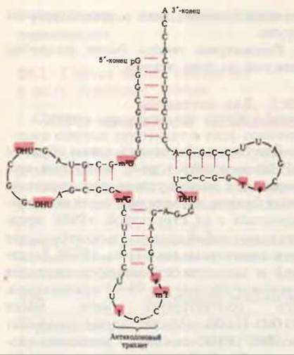
Fig. 29-3. Nucleotide sequence of yeast alanine tRNA, determined by Holley and co-workers and depicted in the form of a cloverleaf. In addition to A, G,
U, and C, the following symbols are used for modified nucleosides: ψ - pseudouridine, I - inosine, T - ribothymidine, DHU - 5,6-dihydrouridine, m2I - 1-methylinosine, m1G - 1-methylguanosine, m2G - N2-dimethylguanosine. Modified nucleosides are highlighted on a red Background. Red lines between parallel segments of the molecule denote complementary Base Pairs. The anticodon has The ability to "recognize" alanine codons in mRNA. Other Structural Features of tRNAs are noted in the text and in Fig. 29-4. In RNA, G can pair with both C and U, although the G-U pair is not as stable as the Watson-Crick G-C pair.
bases. In most tRNAs, a guanylic acid residue (pG) is found at the 5'-end, and the trinucleotide sequence —C—C—A (3') is present at the 3'-end of all tRNAs. If the structural formula of a tRNA is depicted in such a way that the number of intramolecular complementary pairs (i.e., A—U, G—C, and G—U) is maximized, the formula takes the form of a "cloverleaf." Four arms are distinguished in the "cloverleaf"; longer tRNAs additionally contain a short fifth, or extra, arm (Figs. 29-3 and 29-4). Two of these arms are directly involved in the functioning of tRNA as an adaptor. The acceptor arm attaches the specific amino acid (AA), the carboxyl group of which is linked by an ester bond to the 2'- or 3'-hydroxyl group of the 3'-terminal A residue in the tRNA. The anticodon arm contains the anticodon, i.e., a specific triplet of nucleotides that is complementary in the antiparallel direction to the corresponding mRNA triplet (codon) and can form base pairs with it.
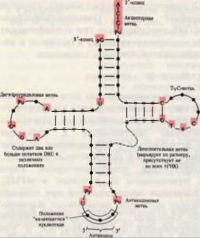
Fig. 29-4. Generalized Secondary structure characteristic of all tRNAs. When depicting the structural formulas of tRNAs taking into account THE PRINCIPLE OF maximizing intramolecular base pairs, all tRNAs adopt a cloverleaf shape. Black circles on the molecular backbone denote nucleoside residues, and red lines are drawn between complementary bases. Positions occupied by the same bases in all tRNAs are highlighted with a red background. tRNA sizes range from 73 to 93 nucleotides. Additional nucleotides are found in the extra and dihydrouridine arms. At the apex of the anticodon arm lies the anticodon loop, which always contains seven unpaired nucleotides. The dihydrouridine arm typically contains up to three DHU residues.
In some tRNAs, the dihydrouridine arm consists of only three hydrogen-bonded base pairs. Designations: R - purine nucleoside, Py - pyrimidine nucleoside, ψ - pseudouridine, G* - guanosine or 2'-O-methylguanosine, T - ribothymidine, DHU - dihydrouridine.
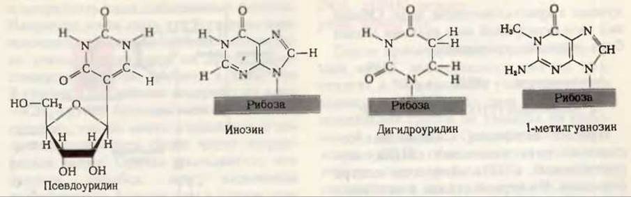
Fig. 29-5. Some of the unusual or modified nucleosides found in tRNA.
Each tRNA has its own distinct anticodon. Its other two main arms are the dihydrouridine arm, which contains the unusual nucleoside dihydrouridine, and the TψC arm; the latter contains the nucleoside ribothymidine (T), which is generally absent in RNA, and the nucleoside pseudouridine (ψ), in which the base and pentose are joined by an unusual carbon-carbon bond (Fig. 29-5).
Yeast phenylalanine tRNA was obtained in crystalline form and subjected to X-Ray Diffraction Analysis, which confirmed that tRNAs obey the principle of maximum intramolecular base pairing via Hydrogen Bonds. However, the three-dimensional structure of tRNA resembles an inverted letter L rather than a cloverleaf (Fig. 29-6). In addition to hydrogen bonds between base pairs, Other types of hydrogen bonds participate in maintaining the Tertiary Structure of tRNA. Because base pairing in RNA is not as strict as in DNA, the paired regions of tRNA lack strict regularity; therefore, unlike the rigid rod-like DNA double helix, tRNA structure is characterized by significant flexibility.
Let us now examine how a specific amino acid is attached to a tRNA molecule by an enzyme.
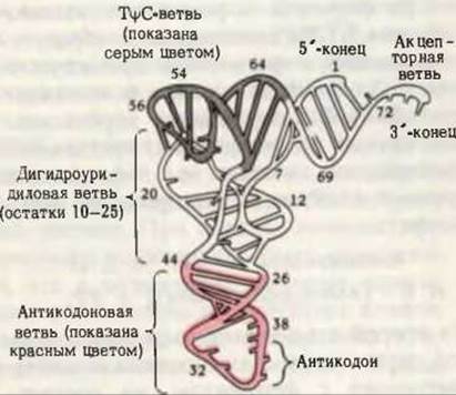
Fig. 29-6. Three-dimensional structure of yeast phenylalanine tRNA determined by X-ray diffraction analysis at 3 Å resolution. It resembles an inverted letter L. [From Kim et al., Science, 185, 436 (1974).]
Last update: 06/08/2026
Editorial and Educational Adaptation: This material has been compiled based on the primary/original source text. The project team performed an editorial review, corrected technical inaccuracies, structured sections, and adapted the content for an educational format.
What was processed:
- elimination of formatting defects (OCR errors, structural breaks, corrupted characters);
- editorial organization of content;
- standardization of terminology in accordance with academic sources;
- verification of factual statements against the original source text.
All mentions of the author, publication year, and origin of the primary text have been preserved in accordance with the source.