BIOCHEMISTRY - Key Regulators and Biological Fluids of the Human Body - 2016
4. CHEMICAL COMPOSITION OF URINE
4.7. Practical Part. Investigation of the Organic Composition and Pathological Components of Urine
Objective: to study the presence of components in the test urine that are present under normal and pathological conditions.
4.7.1 Detection of Organic Components of Urine
Experiment 1. Determination of uric acid in urine
In humans, uric acid is the end product of purine METABOLISM and is an important nitrogenous constituent of urine. Normally, a human excretes an average of 0.6 - 0.7 g of uric acid per day. Its urinary concentration increases when purine-rich foods are included in the diet, as well as in leukemia and febrile conditions. Uric acid is the main component of deposits that form in Cartilage, joints, and tendons in Gout.
Uric acid exists in two tautomeric forms:
Class="center">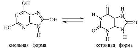
To obtain uric acid crystals, a drop of urine is applied to a Glass slide and carefully evaporated at a Temperature not exceeding 60 - 70 °C by passing the slide over a burner flame and controlling the degree of heating with the back of the hand. A second drop of urine is applied to the same spot and evaporated in the same manner. Add 1 drop of a 1 - 2 % Hydrochloric acid (HCI) solution, cover the slide with a coverslip, and observe The formation of uric acid under a Microscope. Uric acid crystals vary in shape, though rhombic forms predominate. The crystals have a pale yellowish color because they are stained by urinary pigments.
In your report, draw a circle representing the microscope field of view and sketch the observed uric acid crystals inside it.
Experiment 2. Murexide test for uric acid
Place a pinhead-sized amount of uric acid powder onto a porcelain lid, add 1 - 2 drops of concentrated nitric acid (HNO3), and carefully evaporate to dryness without heating above 70 °C, blowing away the evolved lower nitrogen oxide vapors. Allow the brown residue to cool and add a drop of a 25 % ammonia (NH4OH) solution to one edge. A purple-red coloration appears due to the formation of murexide (the ammonium salt of purpuric acid):

Apply 1 drop of a 10 % sodium hydroxide (NaOH) or potassium hydroxide (KOH) solution to the other edge of the brown residue, producing a violet coloration.
In your report, sketch the obtained results using colored pencils and label them accordingly.
4.7.2. Determination of Pathological Components of Urine
4.7.2.1. Detection of Protein in Urine
The urine of a healthy person is virtually protein-free. In certain pathological conditions, protein may appear in the urine in amounts up to 25 g per day (proteinuria, albuminuria).
Color reactions are of little use for detecting protein in urine because both normal and pathological urine contain substances that interfere with the formation of colored reaction products.
To detect protein in urine, three precipitation Methods are commonly used: boiling, Treatment with concentrated nitric acid (Heller's test), and sulfosalicylic acid precipitation.
Experiment 3. Protein precipitation by boiling
First, test the reaction of the urine using litmus paper or a universal indicator. If the urine is acidic, boil 2 - 3 mL of it directly in a test tube; if alkaline, gently acidify it first by dropwise addition of a 1 % acetic acid (CH3COOH) solution until a weakly acidic reaction to litmus is achieved.
In the presence of protein, boiling produces turbidity or a precipitate that does not dissolve upon re-boiling after 3 - 5 drops of a 10 % acetic acid solution are added to the liquid. This distinguishes protein precipitate from the precipitates of calcium and magnesium phosphates and carbonates, which may form upon boiling in a weakly acidic medium but dissolve when heated in a more acidic acetic acid solution.
In your report, indicate the initial pH of the urine. Did a precipitate form upon boiling? If so, did it dissolve upon The addition of acetic acid solution? What Conclusion can be drawn from your observations?
Experiment 4. Heller's test for protein detection
Pour 1 ml of concentrated nitric acid (HNО3) into a test tube and carefully layer 2 - 3 ml of urine down the side of the tube using a pipette. In the presence of protein, an amorphous layer or turbidity—the so-called protein ring—forms at the interface of the two liquids within 2–4 minutes. If protein is absent in the urine, a transparent colored ring appears at the liquid boundary due to the modification of urinary pigments caused by the nitric acid.
In your report, note whether liquid stratification occurred, whether a ring was observed at the phase boundary, and whether it was transparent or turbid. What does this indicate?
Experiment 5. Sulfosalicylic acid test for protein
Pour 1 - 2 ml of urine into a test tube and add 2 - 3 drops of a freshly prepared 20% sulfosalicylic acid solution. If protein is present in the urine, a white precipitate or turbidity forms.
In your report, indicate whether a precipitate formed and what this indicates.
4.7.2.2. Tests for ketone bodies
Acetone bodies, or Ketone Bodies, are products of incomplete fat metabolism, specifically acetone, acetoacetic acid, and 3-hydroxybutyric acid. Under normal fat and Carbohydrate Metabolism, they are almost completely oxidized and are present in the Blood in small amounts (about 0.01 g/L). However, in certain diseases, such as Diabetes Mellitus, as well as during fasting and improper diet, their concentration increases significantly: up to 40 g may be excreted in the urine per day, compared to the normal range of 0.04 - 0.05 g. In such cases, ketone bodies are also excreted through the Skin and Lungs, which can sometimes give the patient's breath an odor of acetone.
Experiment 6. Test for acetone (Liben's test)
The reaction is based on the interaction of acetone with iodine, resulting in the formation of a yellow precipitate of iodoform, which has a characteristic odor.
1. Add 10 drops of acetone, 10 drops of a 10% sodium hydroxide (NаОН) solution, and a few drops of Lugol's solution (1% iodine - 12 solution in a 2% potassium iodide - КI solution) to a test tube.
2. Perform a similar reaction using 2 - 3 ml of urine. If acetone is present in the urine, the liquid becomes turbid.
In your report, describe your observations from the experiments with urine and acetone. Draw a conclusion regarding the presence of acetone in the urine. What does this indicate?
Experiment 7. Test for acetoacetic acid (Gerhardt's test)
The reaction is based on the ability of acetoacetic acid to form a cherry-colored salt with iron(III) chloride solution.
1. Add 10 drops of a 0.5% acetoacetic acid solution (СН3СOOН2СO2Н) and 2 drops of a 10% FеСI3 (iron(III) chloride) solution to a test tube. A cherry-red coloration appears.
2. Perform a similar reaction with urine. Note that after adding the first two drops of the iron(III) chloride solution, a precipitate of iron(III) phosphate (FеРO4) will form, so it is necessary to add a few more drops of this reagent.
In your report, describe your observations from the experiments with acetoacetic acid and urine. Draw a conclusion regarding the presence of acetoacetic acid in the urine and indicate what this signifies.
Last update: 06/08/2026
Editorial and Educational Adaptation: This material has been compiled based on the primary/original source text. The project team performed an editorial review, corrected technical inaccuracies, structured sections, and adapted the content for an educational format.
What was processed:
- elimination of formatting defects (OCR errors, structural breaks, corrupted characters);
- editorial organization of content;
- standardization of terminology in accordance with academic sources;
- verification of factual statements against the original source text.
All mentions of the author, publication year, and origin of the primary text have been preserved in accordance with the source.