Protein Chemistry - Part 2 - Selected Topics in Special Protein Chemistry - Ashmarin I. P. 1968
Contractile proteins of muscle tissue
Contractile proteins within the myofibril. Their isolation and purification
Contractile Proteins are components of myofibrils—thread-like structures 0.5–0.2 μm in diameter and 0.01–0.1 mm in length, which, in turn, make up Muscle fibers. Depending on the type of Muscle tissue, myofibrils vary in the presence or absence of transverse striations. Skeletal Muscles, which comprise the bulk of mammalian musculature (the so-called voluntary musculature responsible for all animal movements and physical work), exhibit transverse striations. In contrast, smooth muscles, found in such structures as the walls of The Stomach, intestines, Blood Vessels, and certain other Internal Organs, appear homogeneous under a Microscope. Only The Heart muscle occupies an intermediate position in A number of features and possesses weakly defined striations.
Contractile proteins exist within the myofibril as a semi-crystalline gel, with a dry residue of 15–20%. The majority of contractile proteins are represented by Myosin (55–60% of the total amount), followed by Actin (typically 20–25%), and finally Tropomyosin (4–30%). The content of the latter is usually low in striated muscles and, conversely, significant in smooth muscles. An uneven distribution of myosin, actin, and tropomyosin along the myofibril has been established. The reason for the transverse striation of skeletal muscles is that these protein substances possess different refractive indices. The Optical Properties of muscle fibers have been studied in detail using phase-contrast and Electron Cell/15.html">Microscopy, as well as X-Ray Diffraction Analysis.
Fig. 3 presents a diagram comparing the elements of myofibril transverse striation observed under an ordinary microscope with data on the localization of contractile proteins. As can be seen, myosin is largely concentrated in the A-band, which exhibits birefringence, or anisotropy. This property is most clearly expressed in the peripheral Zones of the A-band, where myosin is associated with actin. Extraction of myosin from the myofibril with salt solutions leads to the disappearance of anisotropic bands. It is hypothesized that H-bands consist solely of rod-like components of myosin molecules and contain no actin. Optically isotropic I-bands in all directions consist of actin and tropomyosin. The latter is also likely present in the Z-lines, which serve as boundaries for the repeating Structure/83.html">Structural elements of the myofibril, the so-called sarcomeres. The length of a sarcomere is about 2.5 μm, of which about 1.5 μm corresponds to the A-band. The Nature of the so-called M-lines remains unclear.
Class="center">
Fig. 3. Elements of myofibril transverse striation and the probable distribution of contractile proteins within it.
Thick horizontal segments indicate the putative arrangement of myosin molecules, and thin ones indicate actin.
Before examining the composition, structure, and properties of muscle contractile proteins, one should become familiar with the general flowchart for their Isolation and Purification. It is based on the following solubility and stability characteristics of these proteins.
Myosin, as well as the product of its interaction with actin—Actomyosin, is soluble only at a relatively high Ionic strength μ exceeding 0.3 and at pH > 6.5. These features of myosin and actomyosin make it possible, at an ionic strength of 0.1–0.15, to remove readily soluble proteins located predominantly outside the myofibrils in the so-called sarcoplasm, which account for about 25–35% of the total protein of muscle tissue by weight.
The solubility of myosin and, accordingly, the speed and completeness of its extraction from muscle tissue homogenate are greater than those of actomyosin. This circumstance makes it possible, already at The First stage of extraction—by limiting the extraction time from the homogenate to 10–20 min at an ionic strength μ of 0.5 and pH of 6.65–7.0 (0.3 M potassium chloride and 0.15 M potassium phosphate buffer)—to obtain a myosin solution containing negligible (less than 2%) actomyosin impurities. Under these conditions, however, myosin is not extracted completely. Differences in the solubility of myosin and actomyosin are particularly important at an ionic strength close to 0.28, when it is possible to almost completely eliminate actomyosin impurities, which precipitate out under these conditions.
To better separate myosin and actin already at the extraction stage, it is recommended to take into account that at high ionic strength values, ATP causes the dissociation of actomyosin. Therefore, thorough cooling of the initial tissue and its homogenate down to 0–(+4)°C is necessary to prevent the Enzymatic Cleavage of the muscle's own ATP. Cooling is also necessary at subsequent stages of myosin purification, as it is particularly prone to thermal Denaturation. In addition, measures should be taken to prevent premature ATP breakdown in the muscle even before tissue extraction. It must be borne in mind that this occurs during animal spasms at the moment of sacrifice. Therefore, it is best to sacrifice the animal under conditions that exclude spasms (for example, under anesthesia induced by the administration of magnesium chloride).
At the final purification stages, with a gradual decrease in ionic strength to 0.05–0.1, myosin, freed from actin impurities, precipitates from concentrated solutions in the form of pseudo-crystals (crystal-like short fibers visible under a microscope at a magnification of about ×90).
If the researcher's goal is not to separate myosin and actin, but to extract the product of their interaction—actomyosin (so-called myosin B)—then tissue homogenate extraction is carried out at high pH values of 8.0–8.5 and μ = 0.6–0.7 for an extended period—24 hours. Under these conditions, myosin is extracted completely, and actin by approximately 50% (the myosin:actin ratio in actomyosin extracted in this manner is 5:1).
Actin present within muscle tissue is extremely resistant to the denaturing action of such an organic solvent as acetone. Treatment of the tissue homogenate with acetone (before or after myosin extraction), on the one hand, denatures most of the accompanying proteins and, on the other hand, partially extracts lipid components that bind actin in the native muscle tissue, hindering its extraction. Actin is easily extracted with Water from the acetone-treated and dried homogenate, and is then further purified by precipitation from the aqueous extract at its isoelectric point (pH 4.7).
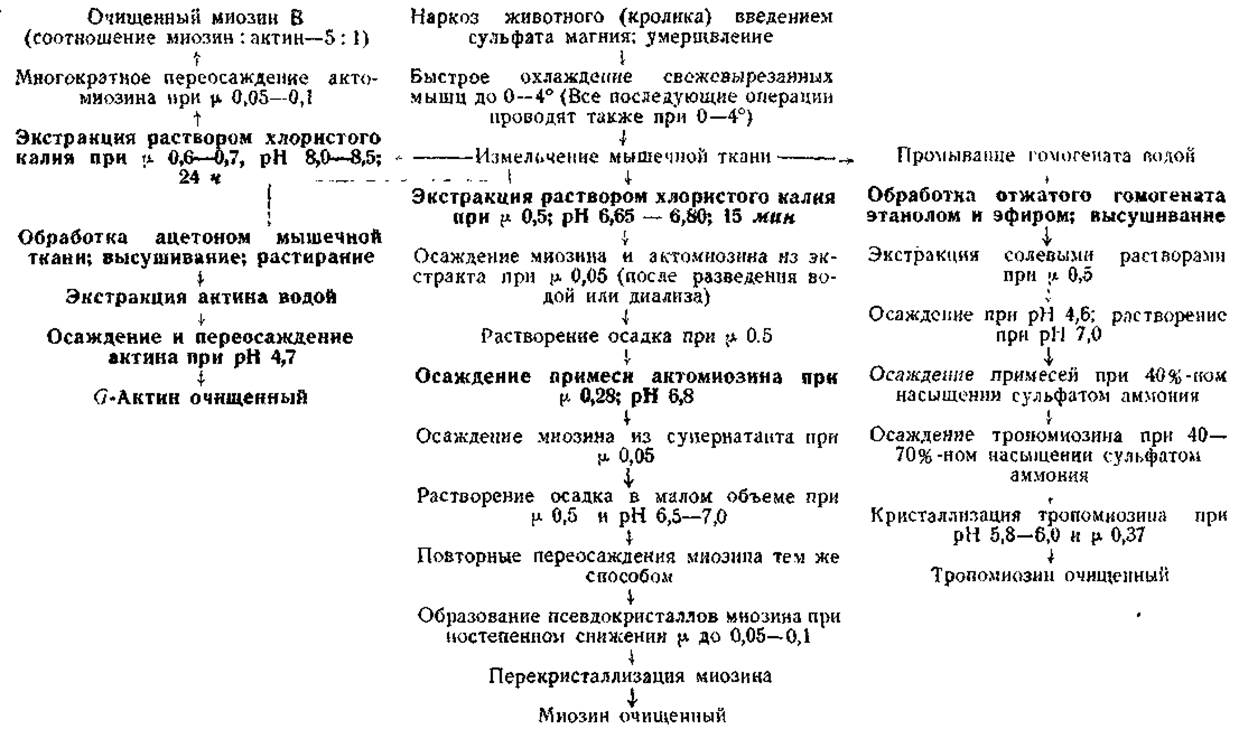
Note. Key steps are highlighted in bold.
Tropomyosin is characterized by extremely high resistance to the action of ethanol and ether. This circumstance (as well as the resistance of actin to acetone) makes it possible to free it from impurities of other proteins during isolation and purification. In addition, treatment of the muscle tissue homogenate with ethanol and ether is necessary to disrupt the bonds between tropomyosin and certain myofibrillar components that hinder its extraction. After this, its complete extraction with water or dilute salt solutions at pH > 6.5 becomes possible. Subsequently, tropomyosin easily crystallizes from concentrated solutions at pH 5.8–6.0 and an ionic strength of 0.37.
The general flowchart for the isolation and purification of myosin, actin, actomyosin, and tropomyosin, based on the Characteristic Properties of proteins listed above, is given on page 20.
Last update: 06/08/2026
Editorial and Educational Adaptation: This material has been compiled based on the primary/original source text. The project team performed an editorial review, corrected technical inaccuracies, structured sections, and adapted the content for an educational format.
What was processed:
- elimination of formatting defects (OCR errors, structural breaks, corrupted characters);
- editorial organization of content;
- standardization of terminology in accordance with academic sources;
- verification of factual statements against the original source text.
All mentions of the author, publication year, and origin of the primary text have been preserved in accordance with the source.