Protein Chemistry - Part 2 - Selected Chapters in Special Protein Chemistry - Ashmarin I. P. 1968
Contractile proteins of muscle tissue
Hypotheses on the mechanisms of muscle contraction
The simplest explanation of Muscle contraction reduces it to the aforementioned reactions of Actomyosin gel with ATP. Once it was demonstrated that actomyosin film threads can perform weight-lifting work in the presence of ATP (see § 5), this reduction at first glance appears entirely justified. Indeed, the complex of the two main Muscle Proteins, by interacting with an energy source that is particularly abundant in muscle, ensures The conversion of chemical energy into mechanical energy. It would seem difficult to demand more conclusive experiments. In reality, however, all experiments of this type merely substantiate the concept that the reaction of actomyosin with ATP is The basis of muscle contraction, but it is not entirely possible to reduce this highly complex process to it alone. As is well known, a characteristic sign of an insufficient understanding of a mechanism is the presence of superfluous details that an inexperienced mechanic discovers with surprise when they believe they have already assembled the system. The same happens when comparing actomyosin models with a myofibril. One can cite many Examples of serious differences both in the Features of the contraction process and in the Structure and COMPOSITION OF THE myofibril versus actomyosin threads. We will dwell only on some of them. For instance, a striking "superfluous detail" is Tropomyosin, which is widely represented in the myofibril but unnecessary for obtaining contractile actomyosin threads. As already indicated in § 6, the putative significance of tropomyosin lies in acting as a locking mechanism for the contracted state. This is presumably achieved through the interaction of rod-like tropomyosin molecules with parallel Actin filaments, when specific Functional groups of both proteins are brought into proximity As a result of contraction.
Recently, evidence has also been obtained indicating that tropomyosin, together with troponin (see § 6), determines the subtle differences between the myofibril and actomyosin in their response to Reagents that specifically bind Calcium Ions. Myofibril contraction is suppressed by a substance such as 1,2-bis(2-biscarboxymethylaminoethoxy)ethane, which forms chelate compounds with calcium ions. This agent has no effect on the superprecipitation of actomyosin precisely because the tropomyosin-troponin complex is absent from its composition.
In general, however, the Functions of tropomyosin are still far from certain. Furthermore, numerous difficulties arise when interpreting the Changes in the electron microscopic appearance of the myofibril that occur during contraction. Above, in Fig. 3 and on page 18, the distribution of muscle proteins in the striated muscle myofibril prior to contraction was described. Characteristically, actomyosin is located there only in the anisotropic A-bands, whereas the H-bands contain Myosin but not actin, while the isotropic I-bands, conversely, contain actin but not myosin. This is believed to be due to the fact that the filamentous myosin and actin molecules lying along the axis of the myofibril overlap only in the A-band. During contraction, a migration of the substance responsible for anisotropy is observed from the A-band into the H-band. The I-band also ceases to be detectable, and the distance between the H- and Z-zones decreases. There are strong grounds to believe that in the process, actin filaments slide along the adjacent myosin molecules and are drawn into the gaps between them within the H-band; from the other end, these same actin filaments pull the Z-bands along with them. As a result, the length of the sarcomere as a whole decreases. What, then, is The Nature of the force that causes the Actin and myosin filaments to move and slide relative to each other? It is believed that this motor is precisely The connection between the H-meromyosin end of the myosin molecule and actin. As indicated in § 3, the reaction with ATP takes place specifically on H-meromyosin (more precisely, on the first subfragment of H-meromyosin, HMM S-1). Apparently, A change in the conformation of H-meromyosin or a change in its bond with actin under METABOLISM/18.html">The Influence of ATP leads to the Displacement of the entire myosin molecule along actin. The Role of the rod-like part of the myosin molecule—L-meromyosin—is to ensure strictly oriented movement along the rod-like actin molecules.
Class="center">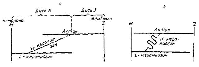
Fig. 11. Schematic diagram illustrating certain elements of Davis's hypothesis. a — relaxation phase b — contraction phase
Overall, the concepts outlined above are shared by many researchers. However, it remains unclear what exactly happens in the region where H-meromyosin contacts actin. It has been suggested that the process is based on the transition of the $\alpha$-structure into folded $\beta$-structures in this region of the myosin molecule. This changes the length of the H-meromyosin bridge between actin and L-meromyosin. It is also possible that after the Introduction of ATP, H-meromyosin dissociates from certain groups and binds to others located at a specific distance from the first. One such hypothesis (Davis's hypothesis) is illustrated by the diagram in Fig. 11.
There are several other "blank spots" in the outlined concepts. However, there is every reason to believe that the coming decade or two will be marked by the complete elucidation of the Molecular Mechanism of muscle contraction, since the fundamental reaction—the Conversion of the chemical energy of ATP into the mechanical energy of actomyosin transformations—has been identified and studied in sufficient detail.
Last update: 06/08/2026
Editorial and Educational Adaptation: This material has been compiled based on the primary/original source text. The project team performed an editorial review, corrected technical inaccuracies, structured sections, and adapted the content for an educational format.
What was processed:
- elimination of formatting defects (OCR errors, structural breaks, corrupted characters);
- editorial organization of content;
- standardization of terminology in accordance with academic sources;
- verification of factual statements against the original source text.
All mentions of the author, publication year, and origin of the primary text have been preserved in accordance with the source.