GENERAL MICROBIOLOGY - T.P. Pyrog - 2004
7. PROKARYOTE SYSTEMATICS
7.6. MODERN TRENDS IN BACTERIAL SYSTEMATICS
7.6.3. Phylogenetic classification
Until 1977, microbiologists generally accepted the existence of two kingdoms of living organisms: PROKARYOTES AND EUKARYOTES. In 1977, C.R. Woese and G.E. Fox compared The nucleotide sequences of 16S rRNA (and analogous eukaryotic 18S rRNA) across a wide range of living organisms. Their results demonstrated
that there are Three kingdoms of living organisms: archaebacteria, eubacteria, and eukaryotes. Archaebacteria and eubacteria differ from each other just as much as each of them differs from eukaryotes.
The results of studies conducted by C.R. Woese and E. Stackebrandt in the first half of the 1980s showed that based on 16S rRNA nucleotide sequence analysis, eubacteria are divided into 11 major groups (Fig. 7.2).
Class="center">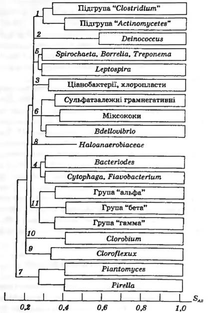
Fig. 7.2. Dendrogram of The formation of major evolutionary lineages within the kingdom Eubacteria
1. Gram-positive Bacteria form a single group (with the exception of Deinococcus), which consists of two subgroups: clostridia (with a low G+C content in DNA) and actinomycetes (with a high G+C content in DNA). This group also includes some Mycoplasmas.
The "Clostridium" subgroup encompasses the following genera: Acetobacterium, Clostridium, Eubacterium, Acetogenium, Acholeplasma, Mycoplasma, Spiroplasma, Bacillus, Brochothrix. Listeria, G a mella, Enters coccus, Streptococcus, Kurthia, Lactobacillus, Leuconostoc, Pediococcus, Peptococcus, PlanococcuSy Staphylococcus, Ruminococcus, Sarcina, Sporolacfobacillus, Sporosarcina, Thermoactinomyces, Erysipelothrix, Aerococcus.
The "Actinomycetes" subgroup includes: Actinomadura, Nocardiopsis, Actinomyces, Bifidobacterium, Propionibacterium, Actinoplanes Ampullariellaу Arthrobacter, Micrococcus, Aureobacterium, Brevibacterium, Cellulomonas, Curtobacterium, Microbacterium, Nocardioides, Kitasatosporia, Microellobosporia, Promicromonosporaу Streptosporangium, Streptoverticillium, Thermomonospora, Corynebacteriumy Dactylosporangium, Micromonospora, Microbacterium, Rhodococcus, Nocardia, Streptomyces, Dermatophilus, Geodermatophilus, Stomatococcus, Frankia.
2. Deinococcus and closely related cocci are characterized by an atypical Cell wall.
3. Cyanobacteria and METABOLISM/14.html">Chloroplasts of Higher Plants and Algae; no heterotrophic representative of this group has been discovered.
4. One of the major eubacterial groups, consisting of the Bacteroides, Flavobacterium, and Cytophaga subgroups. The latter subgroup is divided into two lineages, each containing flavobacteria.
5. Spirochetes form a group of three subgroups combining Spirochaeta, Borrelia, and Treponema, with Leptospira as a peripheral member of this branch.
6. This group unites Bdellovibrio, myxobacteria, and dissimilatory sulfur- and sulfate-reducing bacteria, which share almost no common taxonomic features. The presence of sulfate-reducing bacteria among archaebacteria, Gram-negative, and Gram-positive bacteria indicates that their evolution may have followed different pathways.
7. The group Planctomyces and Pirella comprises budding organisms with a protein cell wall (lacking murein and diaminopimelic acid). A very low SAB value (0.15) compared to other bacteria indicates that members of this group are descendants of the most ancient eubacterial Lineage.
8. Haloanaerobiaceae are anaerobic, moderately halophilic bacteria with a low G+C content in DNA.
9 and 10. Green sulfur bacteria represent a separate evolutionary lineage centered around Chlorobium vibrioforme.
11. The most difficult group to understand and interpret, represented by purple and related Gram-negative bacteria. It includes phenotypically diverse organisms. The composition of group 11 is virtually inconsistent with the existing bacterial Classification given in Bergey's Manual of Determinative Bacteriology, 9th edition. The group combines three main subgroups (alpha, beta, gamma), each containing phototrophic bacteria. Notably, this grouping was established based on both DNA-DNA Hybridization results and 16S rRNA nucleotide sequencing. It is hypothesized that the common ancestor of this bacterial group was a phototroph (Fig. 7.3).
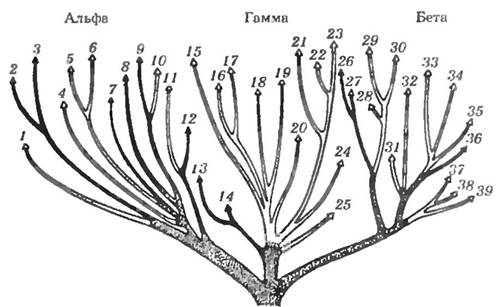
Fig. 7.3. Schematic representation of specific Phylogenetic relationships among genera in the main group of Gram-negative eubacteria. Alpha, beta, and gamma subgroups are indicated. Photosynthetic branches are marked with black lines
In 1988, this group of bacteria was classified into the class Proteobacteria, which currently comprises the provisional subdivisions "alpha", "beta", "gamma", and "delta". The distribution of the most important genera of the class Proteobacteria according to their subdivisions and Nutritional types is presented in Table 7.4.
Table 7.4
Distribution of Proteobacteria by nutritional types
Organotrophs |
Lithotrophs |
Phototrophs |
Alpha subdivision |
||
Acetvbacter, Actdiphilium, Agrobacterium. Аnсatomic robium, Ancylobacter, Ayuaspirillum, Azospirillum. Beijerinckia, Blastobacter, Bradyrhizobium, Brucella, Caulobacter, Cluconobacter. Hyphvmicrobium, Hiphvmonas. Methylobacterium. Paracoccus. Pedumicrobium, Prusthecomic robiam. Stella. Bhizobium. Xanthobacter, Zymomonas |
Nitrobacter |
Rhodobacter, Rhodomic robium. Rhodopila. Rhodopsrudomona. Rhodospinllum |
Beta subdivision |
||
Achmmobnctcr, Alcaligenes, Chrvnwbacterium, Сотатолоя. Derxia, Leptothnx, Xlethylobacter, Methylococcus, Methylomonas, Alethylosinus, Neisllcna, Simonslella. Vitrroscilla |
Nitrosococcus. Nitromlobus, Nttrosospira, Thiobacillus |
Rhodocyclus |
Gamma subdivision |
||
Acinetobacter. Altcromonas, Azvtobactcr, Azomvnas. Citrobacter. Delrya. Enterobacter. Escherichia. Erwinia, Halvmonas, Klebsiella. Legionella, Leucoihrtx. Lysobacter. Moraxclla. Ocranosplrillum, Photobacterium, Proteus, Pseudomonas, Salmonella. Serratia. Vibrio. Xanthomonas |
Beggiatoa, T/nomicrospiru. Thiothnx |
Amoebobacter. Chrvmatium. Ectot hiorhodospira. Lamprocystis, Thiocapsa. Tiodiciion. Thiospirillum |
Delta subdivision |
||
Bdellovibno. Chundrvmyces, Cystobacter. Mухососсus. Pelobacter, Sorangium. Stigmatrlla |
Desulfobacter, Desulfobulbus. Desulfococcus, Desulfonema, Desulfvvtbno, Drsulfuomonas |
|
Different species of this genus fall into different subdivisions
A comparison of the phylogenetic Structure of bacteria with traditional phenotypic systematics (especially group 11) immediately reveals significant differences. A different picture is observed for archaea, whose classification was based on 16S rRNA analysis from the very beginning. The phylogenetic structure of archaea is in complete agreement with their phenotypic classification (Fig. 7.4).
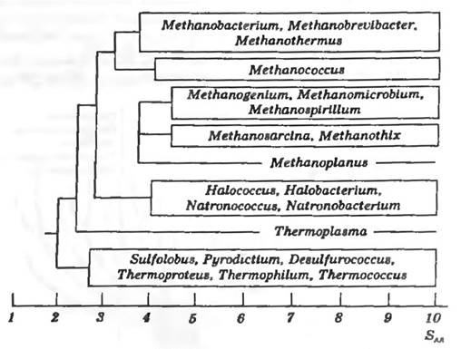
Fig. 7.4. Dendrogram showing the formation of main evolutionary lineages within the archaeal kingdom
In the third and latest edition of The Prokaryotes (1999), unlike the previous ones (1986 and 1992), the Classification of Bacteria is presented taking Phylogenetic Analysis into account. Today, there is an electronic version of this book that is constantly updated and subject to review and revision every four years. Below, for each bacterial group, the number of taxa of various ranks is indicated According to the third edition of The Prokaryotes.
Archaea
Encompasses three orders (Thermoproteales, Sulfolobales, Thermococcales), one family (Halobacteriaceae), two genera (Archaeoglobus and Thermoplasma), and methanogenic bacteria (a taxon of uncertain rank).
Bacteria
1. Division A. Firmicutes includes nine families (Streptomycetaceae, Pseudonocardiaceae, Frankiaceae, Thermomonosporaceae, Streptosporangiaceae, Nocardiaceae, Cellulomonadaceae, Dermatophilaceae, Heliobacteriaceae), 67 genera, as well as taxa of uncertain rank (Lactobacillus, Carnobacterium, anaerobic Gram-positive cocci).
2. Division B. Cyanobacteria — one order (Prochlorales).
3. Division C. Proteobacteria — seven families (Enterobacteriaceae, Vibrionaceae, Pseudomonaceae, Azotobacteriaceae, Halomonadaceae, Chromatiaceae, Ectothiorhodospiraceae), 78 genera, as well as taxa of uncertain rank (Rhizobial, Moraxella, Myxobacteria, manganese-oxidizing bacteria, lithotrophic ammonium-oxidizing bacteria, unusually curved bacteria, Gram-negative mesophilic sulfate-reducing bacteria).
4. Spirochetes — four genera (Spirochaeta, Treponema, Borrelia, Leptospira).
5. Chlorobiaceae — order Cytophagales, family Chlorobiaceae, seven genera.
6. Chlamydiae — genus Chlamydia.
7. Planctomycetes and related bacteria — order Plantomycetales and four genera.
8. Family Deinococcaceae, genus Thermus, and related microorganisms.
9. Chloroflexaceae and related bacteria — family Chloroflexaceae and four genera.
10. Genus Verrucomicrobium.
11. Order Thermotogales.
Some taxa remain phylogenetically unassigned. These include most forms that grow in Symbiosis with other organisms; thermophilic aerobic hydrogen-oxidizing bacteria; morphologically diverse sulfur-oxidizing bacteria; large symbiotic spirochetes; mycoplasma-like organisms (pathogenic to plants); 12 bacterial genera (including Zoogloea, Leptotrichia, Gallionella, Caulococcust Kuznezovia, Siderocapsa, Fusobacterium), and several others.
A comparative evaluation of the phylogenetic classification of bacteria presented in The Prokaryotes (1999) and the phenotypic classification in Bergey's Manual of Determinative Bacteriology, 9th edition, reveals substantial differences between them.
It should be noted that the tenth edition of Bergey's Manual of Systematic Bacteriology is currently in preparation, which will incorporate the latest advances in bacteriology and Taxonomy. This new edition of one of the most authoritative guides to bacterial taxonomy has been completely reorganized to reflect the phylogenetic classification of bacteria. Compared to the previous edition of Bergey's Manual of Systematic Bacteriology, published in 1984, this edition has been expanded with descriptions of over 2,200 new species and 390 new genera. The tenth edition of Bergey's Manual of Systematic Bacteriology will consist of five volumes. The first volume is dedicated to archaea and phototrophic bacteria, the second to Gram-negative bacteria (The Proteobacteria), the third to Gram-positive bacteria with low DNA G+C content, the fourth to Gram-positive bacteria with high DNA G+C content, and the fifth to planctomycetes, spirochetes, fusobacteria, etc.
Last update: 13/08/2026
Editorial and Educational Adaptation: This material has been compiled based on the primary/original source text. The project team performed an editorial review, corrected technical inaccuracies, structured sections, and adapted the content for an educational format.
What was processed:
- elimination of formatting defects (OCR errors, structural breaks, corrupted characters);
- editorial organization of content;
- standardization of terminology in accordance with academic sources;
- verification of factual statements against the original source text.
All mentions of the author, publication year, and origin of the primary text have been preserved in accordance with the source.