Orthopedics - Oleksa A.P. 2006
Neurogenic arthropathies
Chondromatosis of the joint
Joint chondromatosis is a relatively rare condition that predominantly affects working-age men of athletic build. It occurs three times less frequently in women.
This condition was first described by Henderson and Jones, which is why it is also known by their names.
The Essence of the disease lies in the presence of cartilaginous or osteocartilaginous bodies (varying in small size) within the synovial membrane, as well as loose bodies within the joint cavity that have detached from this membrane.
Typically, chondromatosis localizes in the knee or elbow joints, and very rarely in the hip and shoulder joints (Fig. 365).
The appearance of loose cartilaginous and osteocartilaginous bodies in the joint cavity can occur in arthrosis, osteochondritis dissecans, neuropathic arthropathies, etc.; however, these differ from primary chondromatosis, the Etiology of which remains ultimately unclear to this day.
There are many hypotheses regarding the etiology of joint chondromatosis, but it is generally accepted to be a consequence of metaplasia in the inner Connective Tissue layer of the synovial membrane. Histological examinations of the synovial membrane in chondromatosis reveal clusters of swollen connective tissue Cells, indicating their metaplasia into Cartilage. Once The formation of small cartilage islets begins within the synovial membrane, they enlarge very slowly, becoming entirely enveloped by this membrane. Due to joint movements, these cartilage islets initially remain attached to the membrane by a stalk, which subsequently tears off, leaving the cartilaginous bodies freely floating within the joint cavity.
Class="center">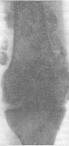
Fig. 365. Radiograph of the elbow joint with chondromatosis and loose chondromatous bodies removed during surgery.
The detached loose cartilaginous bodies float in the synovial fluid and grow by absorbing nutrients from it. This is why surgery often reveals bodies of various sizes, sometimes reaching the size of a bean. They are covered with a smooth layer of cartilage, and cross-sectioning shows that they are ossified in the center. Early signs of ossification can also be detected histologically in smaller loose cartilaginous bodies.
Initially, the disease is clinically asymptomatic, but over time, as loose bodies form in the joint cavity, the patient may experience crepitus and a aching pain in the joint during ambulation, sometimes accompanied by joint Swelling due to synovitis. Larger bodies can become trapped between the articular surfaces during movement and lock the joint. Frequent locking episodes are the primary reason patients seek medical attention.
Physical examination may reveal smoothed contours of the knee or elbow joint; Palpation during movement reveals joint crepitus and even mobile, evanescent loose bodies.
Radiographs reveal only calcified or ossified loose cartilaginous bodies, which can be numerous and of varying sizes. They may be located not only within the joint cavity but also in communicating recesses that are significantly expanded due to synovitis (such as the upper suprapatellar pouch of the knee or the lower pouch of the shoulder joint, etc.).
Long-standing chondromatosis may lead to The Development of secondary arthrosis in the affected joint.
To visualize non-ossified loose cartilaginous bodies within the joint, contrast arthrography or sonography is performed.
Radiological findings in chondromatosis are so characteristic that Differential Diagnosis with other joint pathologies is generally unnecessary.
The Treatment for joint chondromatosis is exclusively surgical. The Procedure is performed under general anesthesia and using a hemostatic tourniquet.
The joint cavity is exposed using a classical surgical approach. Loose cartilaginous bodies immediately escape from the wound along with the synovial fluid. After removing all loose bodies, the joint is irrigated with a normal saline solution or a 1:5000 furatsilin (nitrofurazone) solution, and the synovial membrane is inspected. Occasionally, small hypertrophied villi with visible white inclusions can be detected within it. Considering the pathogenetic nature of chondromatosis, regardless of whether pathological Changes in the synovial membrane are macroscopically apparent, synovectomy or the technically simpler synovial capsulectomy must be performed.
The wound is closed in layers, and the joint is drained for 24 hours. The limb is immobilized with a plaster splint until the wound heals.
To prevent scarring contracture following synovectomy, it is advisable to intra-articularly administer a 0.5% novocaine solution two to three days postoperatively, which provides analgesia and prevents adhesion formation in the operated deep tissue layers. Sometimes, surgeons limit the procedure to the removal of loose bodies and partial synovectomy using arthroscopic techniques.
Beginning on the fifth or sixth day, the patient commences mobilization exercises for the operated joint.
Following the removal of cartilaginous bodies from the joint and synovectomy, functional outcomes are almost always favorable.
Last update: 10/08/2026
Editorial and Educational Adaptation: This material has been compiled based on the primary/original source text. The project team performed an editorial review, corrected technical inaccuracies, structured sections, and adapted the content for an educational format.
What was processed:
- elimination of formatting defects (OCR errors, structural breaks, corrupted characters);
- editorial organization of content;
- standardization of terminology in accordance with academic sources;
- verification of factual statements against the original source text.
All mentions of the author, publication year, and origin of the primary text have been preserved in accordance with the source.