Human Anatomy - Kotsan I. Y. 2009
Skeleton of the Lower Limb
Joints of the Bones of the Lower Limb Girdle
The BONES OF THE Pelvic Girdle are connected by almost all Types of bone joints: discontinuous joints (sacroiliac joint), semi-discontinuous joints (Pubic Symphysis), and continuous joints—syndesmoses (ligaments), synchondroses (cartilages between individual PARTS OF THE hip bone), and synostoses (after the fusion of these parts into a single hip bone).
The sacroiliac joint (articulatio sacroiliaca) is formed by the auricular surfaces of the sacrum and ilium. The articular surfaces on both bones are covered with a layer of fibrocartilage, which is thicker on the sacrum. The joint is simple and flat in shape. Movements occur around all three axes of rotation, but their range is minimal (only 3—5°), primarily serving a Shock-absorbing function. The Joint Capsule is tightly stretched and very strong. It fuses with the periosteum of the sacrum and hip bone, as well as with the ligaments that reinforce this joint. Anteriorly, the joint capsule is reinforced by the anterior (ventral) sacroiliac ligaments (ligg. sacroiliaca anteriora (ventralia)), which extend from the pelvic surface of the sacrum to the ilium (Fig. 76). On the posterior surface of the joint are the interosseous sacroiliac ligaments (ligg. sacroiliaca interossea) and posterior (dorsal) sacroiliac ligaments (ligg. sacroiliaca posteriora (dorsalia)) (Fig. 77). The interosseous ligaments are the strongest ligaments of the sacroiliac joint. They fuse with the posterior surface of the joint capsule, filling the narrow cleft between the sacral and iliac tuberosities and attaching to them. The posterior (dorsal) sacroiliac ligaments originate from the upper and lower posterior iliac spines and attach to the lateral crest of the sacrum.
Class="center">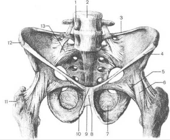
Fig. 76. Ligaments of the pelvis and hip joint (anterior view)
1 — fourth lumbar vertebra, 2 — anterior longitudinal ligament, 3 — iliolumbar ligament, 4 — inguinal ligament, 5 — joint capsule, 6 — iliofemoral ligament, 7 — obturator membrane, 8 — pubic symphysis, 9 — arcuate ligament of the pubis, 10 — superior pubic ligament, 11 — greater trochanter, 12 — anterior superior iliac spine, 13 — anterior sacroiliac ligament

Fig. 77. Ligaments of the pelvis and hip joint (posterior view)
1 — iliolumbar ligament, 2, 3 — dorsal sacroiliac ligaments, 4 — iliofemoral ligament, 5 — ischiofemoral ligament, 6 — joint capsule, 7 — sacrotuberous ligament, 8 — lateral sacrococcygeal ligament, 9 — posterior superficial sacrococcygeal ligament, 10 — falciform process, 11 — sacrotuberous ligament (cut), 12 — circular zone, 13 — acetabular labrum, 14 — obturator membrane, 15 — sacrospinous ligament, 16 — dorsal sacroiliac ligament (sacrotuberous ligament cut), 17 — posterior superior iliac spine
The sacroiliac joint is additionally reinforced by ligaments located at some distance from it and attached to adjacent bones:
— iliolumbar ligament (lig. iliolumbale), which originates from the transverse processes of the two lower lumbar vertebrae and attaches to the iliac crest and its tuberosity;
— sacrospinous ligament (lig. sacrospinale), which originates from the ischial spine and terminates inferiorly on the lateral part of the sacrum and the lateral surface of the coccyx;
— sacrotuberous ligament (lig. sacrotuberale), which originates from the ischial tuberosity of the ischium, fans out, and attaches to the lateral margins of the sacrum and coccyx. The continuation of the sacrotuberous ligament anteriorly and inferiorly is its falciform process (processus falciformis). The sacrotuberous and sacrospinous ligaments, together with the greater and lesser sciatic notches, bound two openings: the greater sciatic foramen (foramen ishiadicum majus) and the lesser sciatic foramen (foramen ishiadicum minus), through which Muscles, Blood Vessels, and nerves emerge from the pelvis.
The pubic symphysis (symphysis pubica) is formed by the opposing symphyseal surfaces of the pubic bones, which are covered with hyaline Cartilage and connected by a fibrocartilaginous plate—the interpubic disc (discus interpubicus). The pubic symphysis belongs to the transitional type of bone joints known as symphyses (hemizygous or semi-joints), since the center of the disc contains a small, sagittally oriented cleft-like cavity that appears In the second year of life, transforming the synchondrosis into a hemijoint. Among all bone joints, the pubic symphysis exhibits the most pronounced Sexual Dimorphism in Structure. In males, the cartilaginous disc is thinner, stronger, and vertically longer. In females, it is softer, thicker, and shorter. The cavity inside the cartilage is larger in females, particularly during Pregnancy, when the cartilage softens and the cleft widens, causing the symphysis to expand and enlarge the pelvic cavity, which is of great significance during childbirth.
The pubic symphysis is reinforced by two transversely oriented ligaments: superiorly by the superior pubic ligament (lig. pubicum superius), and inferiorly by the arcuate ligament of the pubis (lig. arcuatum pubis) (Fig. 76).
The obturator membrane (membrana obturatoria), which Functions as an intrinsic ligament of the hip bone, is stretched across the margin of the obturator foramen. Spanning across the obturator groove, it forms—together with this groove and the obturator tubercles—the obturator canal (canalis obturatorius), through which pass the vessels and nerve of the same name.
Last update: 08/08/2026
Editorial and Educational Adaptation: This material has been compiled based on the primary/original source text. The project team performed an editorial review, corrected technical inaccuracies, structured sections, and adapted the content for an educational format.
What was processed:
- elimination of formatting defects (OCR errors, structural breaks, corrupted characters);
- editorial organization of content;
- standardization of terminology in accordance with academic sources;
- verification of factual statements against the original source text.
All mentions of the author, publication year, and origin of the primary text have been preserved in accordance with the source.