Human Anatomy (with the Fundamentals of Dynamic and Sports Morphology) - Ivanitsky M. F. 2008
The Doctrine of Internal Secretion Organs
Parathyroid Glands
The Parathyroid glands (Fig. 145), four in number, are located on the posterior surface of the thyroid lobes along the course of the inferior thyroid artery. They usually lie somewhat asynchronously, two on each side of the gland, and occasionally within the glandular tissue itself. Each parathyroid gland measures 4–8 mm in length, 3–4 mm in width, and 2–3 mm in thickness. They consist of cords or clusters of epithelial Cells known as parathyroidocytes. Externally, they are covered by a Connective Tissue capsule from which delicate septa extend into the organ, separating the trabeculae from one another.
The parathyroidocytes of the parathyroid glands secrete parathormone, a hormone that promotes calcium assimilation and deposition in the Bone tissue, while enhancing renal excretion of phosphorus. The removal of these glands significantly increases the excitability of the neuromuscular apparatus—an effect so profound that it can trigger not only Muscle twitching, but also severe spasms and even death.
Class="center">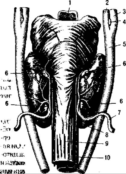
Fig. 145. Parathyroid glands:
1 — epiglottis; 2 — external carotid a.; 3 — internal carotid a.; 4 — carotid body; 5 — common carotid a.; 6 — parathyroid glands; 7 — inferior thyroid a.; 8 — vagus n.; 9 — Esophagus; 10 — Trachea (after Spalteholz)
Last update: 08/08/2026
Editorial and Educational Adaptation: This material has been compiled based on the primary/original source text. The project team performed an editorial review, corrected technical inaccuracies, structured sections, and adapted the content for an educational format.
What was processed:
- elimination of formatting defects (OCR errors, structural breaks, corrupted characters);
- editorial organization of content;
- standardization of terminology in accordance with academic sources;
- verification of factual statements against the original source text.
All mentions of the author, publication year, and origin of the primary text have been preserved in accordance with the source.